Chapter 8 Lens
INTRODUCTION
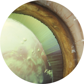
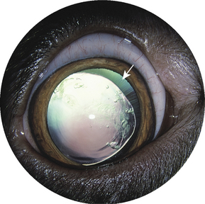
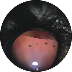
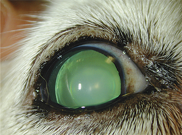
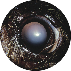
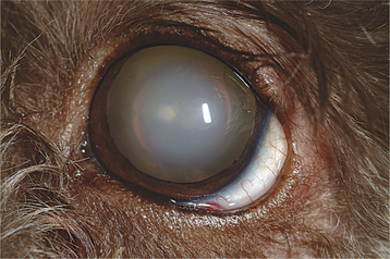
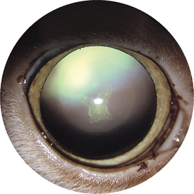
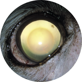
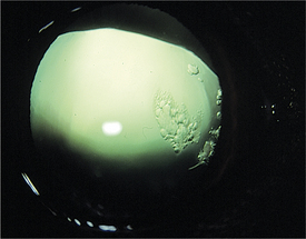
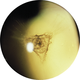
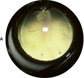
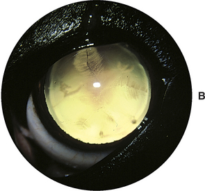
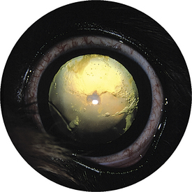
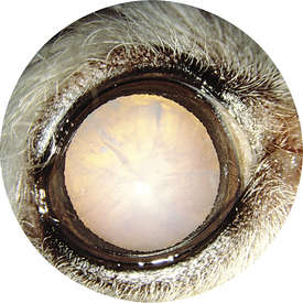
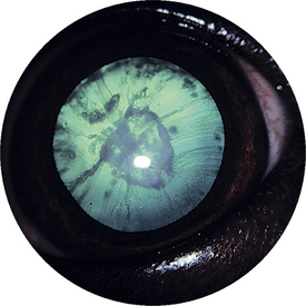
Figure 8-15 Incipient cataracts (vacuoles) positioned equatorially in a dog with hypertensive retinopathy.
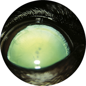
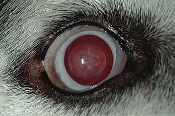
Figure 8-16 Diffuse posterior cataracts with peripheral equatorial vacuoles in a dog with diabetes mellitus.
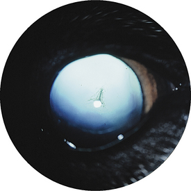
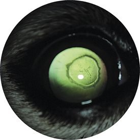
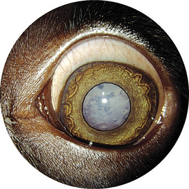
Figure 8-19 Incipient cataract in the lens periphery associated with progressive retinal atrophy (PRA) in a dog.

< div class='tao-gold-member'>
Only gold members can continue reading. Log In or Register to continue
Stay updated, free articles. Join our Telegram channel

Full access? Get Clinical Tree


