6 Large eyelid mass
PRESENTING SIGNS
Dogs, and less frequently cats, will be presented due to a change in appearance and possibly a red eye. There is also likely to be some ocular discharge. The onset is often gradual – the owner has probably been aware of a problem for a few weeks but the lesion increases in size such that they present the patient. Alternatively, the lid mass might have been present for months but has suddenly changed – growing rapidly, bleeding or itching such that the patient rubs and further traumatizes the lesion. No other symptoms are generally reported. Most patients are older adults (8 years and over) and no breed or sex predilection is apparent with the exception of squamous cell carcinoma which is more common in white furred animals, especially cats.
CASE HISTORY
In general there is little in the way of previous history. However, some dogs will have had an eyelid mass or other skin masses in the past, which frequently have been removed, often without histopathological examination. In some cases regrowth can occur very quickly – within weeks – of the previous excision. In other examples owners initially suspect that the current problem is similar to small benign warty growths from which the dog suffers, but the lesion either suddenly enlarges quickly, or changes in appearance – becomes friable and starts to bleed, or causes the pet to rub for example – and then they present the animal.
CLINICAL EXAMINATION
General clinical examination is normally unremarkable, although occasionally a localized lymphadenopathy is present and should be checked for. On ophthalmic examination a large mass can be seen on either the upper or lower lid margin. The periorbital skin might be thickened and inflamed. The mass itself will probably involve the lid margin, but not invariably so (Figure 6.1). The mass might be smooth or lobulated and can vary in colour from pale pink through red to darkly pigmented. The palpebral conjunctiva will be inflamed and the lid margin should always be everted to check the internal extent of the mass – frequently what can be seen from the outside is just the ‘tip of the iceberg’ (Figure 6.2). Some ocular discharge is normally present – this might be serous but is commonly mucopurulent. In some patients it will be excessive and can stick the eyelids together. Schirmer tear test is usually normal. The cornea should be carefully examined – some keratitis is common where the lid mass rubs on the cornea during blinking, and ulceration is not uncommon. Intraocular examination is usually unremarkable, although there might be slight miosis, representing a reflex uveitis if significant trigeminal pain is present.

Figure 6.1 Lid mass affecting the palpebral conjunctiva rather than the lid margin which presented as a variable lid swelling rather than an obvious mass. Mast cell tumour.

Figure 6.2 Upper eyelid mass in a 10-year-old terrier. The grey papillomatous growth at the lid margin was all that could be seen with the lid in its normal position, but on everting it a much larger pink lesion can be seen extending both medially and posteriorly.
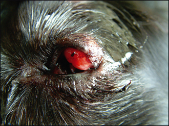
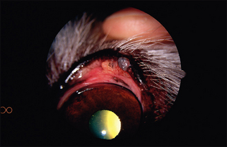
Table 6.1 Eyelid tumour types in dogs
Tumour type | Typical appearance |
---|---|
Papilloma | |
Adenoma/adenocarcinoma | |
Histiocytoma | |
Melanoma | |
Mast cell tumour |
![]() Stay updated, free articles. Join our Telegram channel![]() Full access? Get Clinical Tree![]() ![]() ![]() |