CHAPTER | 12 Keratinization and Seborrheic Disorders


Callus
Diagnosis
Treatment and Prognosis
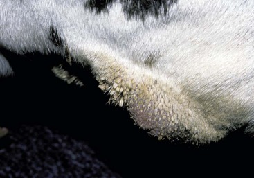
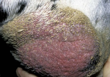
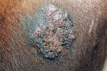
FIGURE 12-2 Callus.
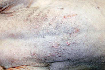
Close-up of the lesion in Figure 12-1. The large alopecic area of thickened skin over the elbow is typical of this syndrome. Often in short-coated dogs, the hairs become impacted within the follicles and callus.
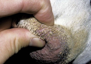
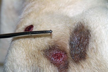
FIGURE 12-3 Callus.
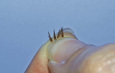
Close-up of the lesion in Figure 12-1. The clinician is gently squeezing the callus to express the impacted hairs, which are now exuding from the surface of the skin. These hairs serve as a nidus for recurrent infection.
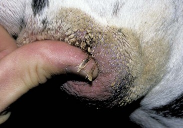
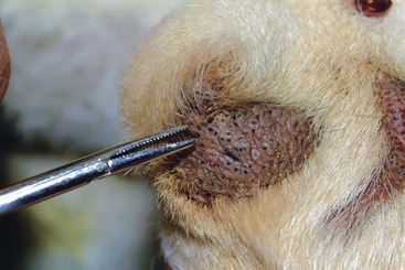
FIGURE 12-4 Callus.
Close-up of the lesion in Figure 12-1. The exuded hairs are apparent. This technique is not recommended because forcing the hairs to rupture internally could result in cellulitis and scarring.
Feline Acne
Treatment and Prognosis


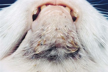
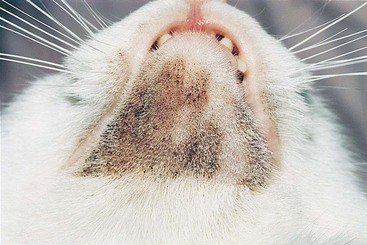
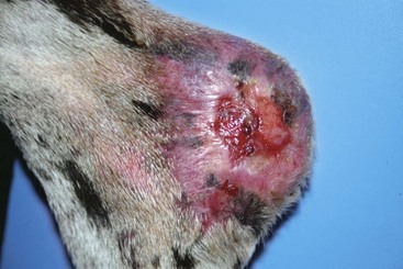
FIGURE 12-19 Feline Acne.
Alopecia and scarring remained as sequelae after treatment with topical mupirocin ointment.
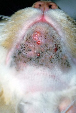
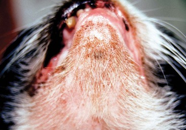
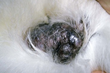
FIGURE 12-21 Feline Acne.
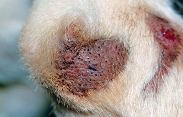
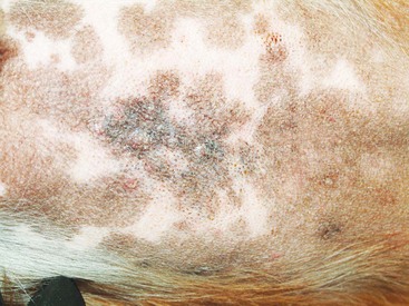
The brown discoloration is typical of comedo formation associated with feline acne.
Idiopathic Nasodigital Hyperkeratosis
Treatment and Prognosis


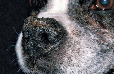
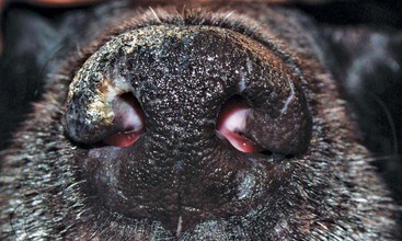
FIGURE 12-23 Idiopathic Nasodigital Hyperkeratosis.
Severe frondlike hyperkeratotic projections with crust formation on the nose of an old Boxer.
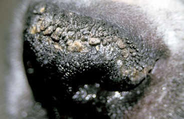
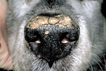
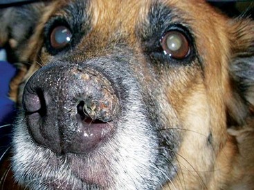
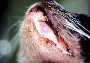
FIGURE 12-25 Idiopathic Nasodigital Hyperkeratosis.
Thick, adherent crusts cover most of the nasal planum in this dog.
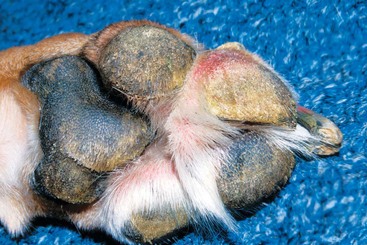
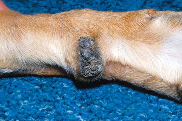
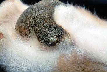
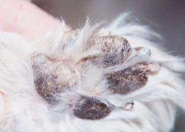
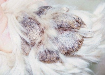
FIGURE 12-28 Idiopathic Nasodigital Hyperkeratosis.
A focal area of hyperkeratosis on the central pad of a Greyhound (Greyhound corns).
Hereditary Nasal Parakeratosis of Labrador Retrievers
Diagnosis
Treatment and Prognosis
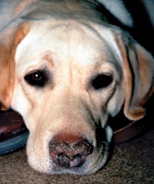
FIGURE 12-31 Hereditary Nasal Parakeratosis of Labrador Retrievers.
Hyperkeratosis and crusting on the nose of a young adult Labrador.
(Courtesy M. Paradis.)

Parasympathetic Nasal Hyperkeratosis (xeromycteria: dry nose)
Diagnosis
Treatment and Prognosis


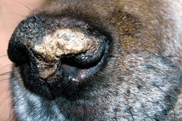
FIGURE 12-33 Parasympathetic Nasal Hyperkeratosis.
Hyperkeratosis and crusting asymmetrically affecting the nasal planum.
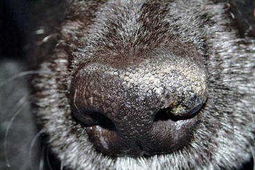
FIGURE 12-34 Parasympathetic Nasal Hyperkeratosis.
Close-up of the dog in Figure 12-33. The asymmetrical (only half) pattern of crusting is apparent.