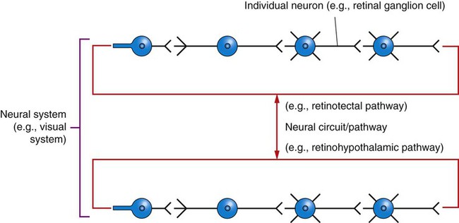
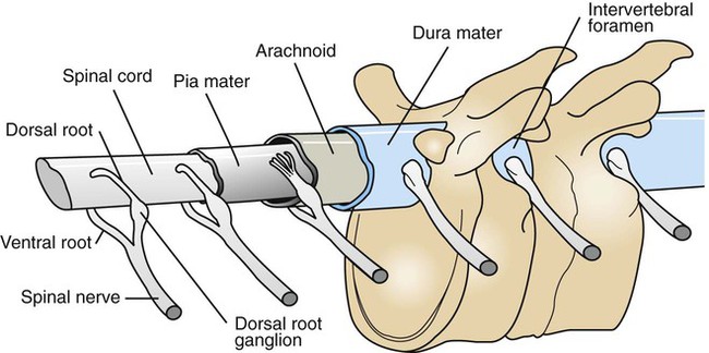
Introduction to the Nervous System


Introduction to the Nervous System


The nervous system is the first multicellular system described in this book because it is one of the major coordinating systems of the body, and because clarifying many of the concepts that concern the nervous system is important for understanding other systems of the body.


Most clinical signs in veterinary neurology involve abnormal movement (e.g., seizures, paralysis); therefore the physiology of muscle control, posture, and locomotion is emphasized in the following chapters. Because veterinary ophthalmology is an extensive subspecialty, the physiology of vision is also covered. Other sensory systems that can produce easily recognizable clinical signs (e.g., vestibular system, hearing) are discussed in Section II as well. Understanding the autonomic nervous system is essential for understanding pharmacology and the involuntary control of many of the body’s most critical functions. Similarly, understanding the blood-brain barrier and the cerebrospinal fluid system is essential to understanding the results of the diagnostic cerebrospinal fluid tap and the homeostasis of the cellular microenvironment of the central nervous system. The electroencephalogram and sensory-evoked potentials are described because of their clinical importance in veterinary medicine. Because of space limitations, only the basic physiological concepts essential to understanding the mechanisms of disease and the practice of veterinary medicine are emphasized. For a more expansive study of neurophysiology, the reader should refer to the texts listed in the chapter bibliographies.


The Neuron Is the Major Functional Unit of the Nervous System


The major functional unit of the nervous system is the neuron, a cell type whose shape varies considerably with its location in the nervous system. Almost all neurons have an information-receiving area of the cell membrane, usually called the dendrite; a cell body, or soma, containing the organelles for most cell metabolic activity; an information-carrying extension of the cell membrane, called an axon; and a presynaptic terminal at the end of the axon to transmit information to other cells. The axon is often covered with a fatty coating called the myelin sheath that enhances the speed of information transfer along the axon’s length.


Neurons do not exist in isolation; they are usually interconnected within neural circuits or pathways that serve a specific function (Figure 3-1). Neural circuits/pathways that are related in function are often collectively referred to as neural systems. For example, the retinotectal pathway provides information for reflex orientation of the eyes to the position of a light source, whereas the retinohypothalamic pathway carries information affecting the body’s physiological rhythms in response to light-dark cycles. These individual neural pathways are both part of the visual system.


The other cell type in the nervous system is the glial cell. Glial cells play important roles in producing the myelin sheaths of axons, modulating the growth of developing or damaged neurons, buffering extracellular concentrations of potassium and neurotransmitters, formation of contacts between neurons (synapses), and they participate in certain immune responses of the nervous system. Glial cells do not produce action potentials, but growing evidence indicates that they can indirectly monitor the electrical activity of neurons and use this information to modulate the effectiveness of neural communication. However, not all glial actions are beneficial to the nervous system. Glial-mediated neuroinflammatory responses have been implicated in some neurodegenerative diseases and in the development of chronic pain conditions.


The Mammalian Nervous System Has Two Major Subdivisions: the Central Nervous System and the Peripheral Nervous System


The central nervous system (CNS) is divided into the brain and spinal cord (Box 3-1). A series of protective bones surround the entire CNS. The brain is surrounded by the skull, and the spinal cord is surrounded by a series of cervical, thoracic, and lumbar vertebrae and ligaments.


The peripheral nervous system (PNS) is composed of the spinal and cranial nerves that carry electrical signals, called action potentials, away from or toward the CNS. These nerves are bundles of PNS axons. The axons carrying action potentials toward the CNS are called afferents, and those carrying such signals away are efferents. One way to group the elements of the PNS functionally is into sensory and motor subsystems. The elements of spinal and cranial nerves that serve a motor function are (1) axons of somatic efferent neurons, which carry action potential commands from the CNS to junctions, called synapses, at skeletal muscles, and (2) axons of visceral efferent neurons, which carry action potentials toward synapses with peripheral neurons that control smooth muscle, cardiac muscle, and some glands. PNS components serving a sensory function are axons of afferent neurons that bring action potential messages to the CNS from peripheral sensory receptors. These receptors are directly or indirectly responsible for transducing energy from the body’s external or internal environment into action potentials that travel to the CNS. The intensity of this energy’s stimulation of the receptor is encoded by changing the frequency of action potentials as the intensity of stimulation changes.


Spinal and cranial nerve sensory components are axons of (1) somatic afferent neurons and (2) visceral afferent neurons. Somatic afferent axons carry action potentials resulting from stimulation of receptors such as the photoreceptors of the eye, auditory receptors of the ear, and stretch receptors of the skeletal muscle. Action potentials generated by stretch receptors or chemoreceptors (e.g., O2, CO2) located within visceral organs of the chest and abdomen are carried to the CNS along visceral afferent axons. Visceral efferent and afferent axons are part of the autonomic nervous system; the portions of the PNS and CNS responsible for involuntary control of smooth muscle, cardiac muscle, some glands, and many physiological life support functions (e.g., heart rate, blood pressure, digestion).


Peripheral nerve axons converge to form a single spinal nerve at each of the intervertebral foramina. Within the spinal canal, afferent sensory and efferent motor axons are separated; afferent sensory axons enter the spinal cord through the dorsal roots, whereas the efferent motor axons exit the spinal cord through the ventral roots (Figure 3-2).


< div class='tao-gold-member'>

Stay updated, free articles. Join our Telegram channel

Jul 18, 2016 | Posted by in PHARMACOLOGY, TOXICOLOGY & THERAPEUTICS | Comments Off on Introduction to the Nervous System

Full access? Get Clinical Tree

Get Clinical Tree app for offline access