12 Introduction to crusting and scaling
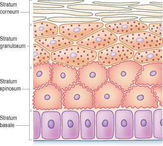
The outermost layer of the skin, the epidermis, is made up of multiple layers of cells (Fig. 12.1). The predominant cell type is the keratinocyte, and the epidermis is divided into basal, spinous, granular (variably present in dogs and cats) and cornified layers depending on the morphological features keratinocytes assume as they undergo progressive differentiation to form the stratum corneum.
SCALING
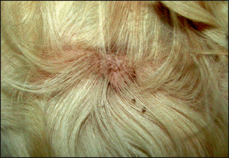
Many diseases affect the normal maturation, differentiation and desquamation processes, and can result in scaling. The appearance and distribution of the scale varies depending on the causative disease. Diffuse dorsal scaling is seen in association with pruritus as a result of cheyletiellosis (see Chapter 5). Another common cause of scaling with a multifocal distribution is as a result of epidermal collarette formation. These are circular rims of scale that are the remains of pustules after they have ruptured (Fig. 12.2). Diseases associated with epidermal collarettes include pyoderma, demodicosis, dermatophytosis and pemphigus foliaceus. Multifocal patches of scaling evident over the dorsal trunk commonly arise from sites of pyoderma and are due to the formation of epidermal collarettes. Tightly adherent patches of scale which are difficult to remove are seen in some forms of ichthyosis, a rare, congenital disorder occurring mainly in young dogs associated with a failure of breakdown of intercorneocyte adhesion (Fig. 12.3). Follicular casts, nit-like accumulations of scale surrounding hair shafts, are representative of hair follicle pathology and are seen in follicular diseases such as sebaceous adenitis and vitamin A-responsive dermatitis (Fig. 12.4).
Stay updated, free articles. Join our Telegram channel

Full access? Get Clinical Tree


