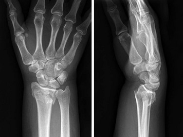
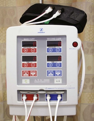
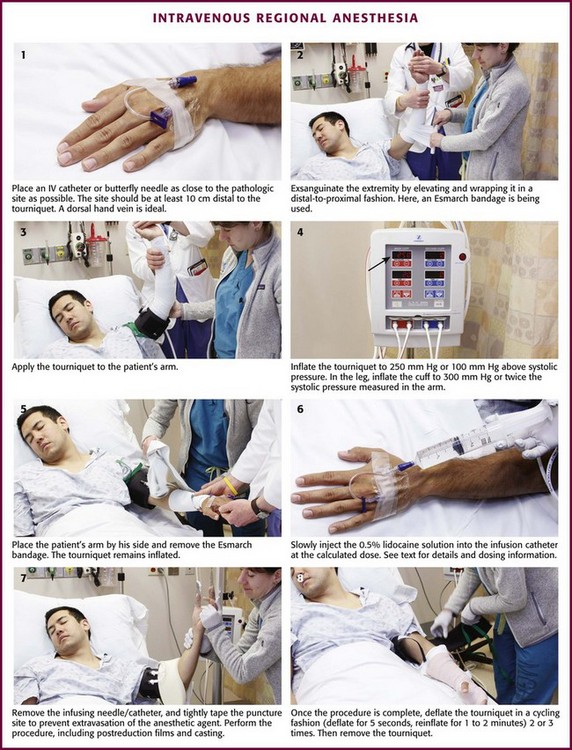
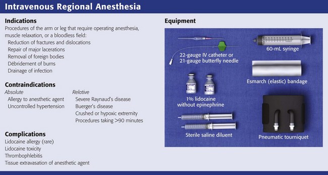
Chapter 32 Review Box 32-1 Intravenous regional anesthesia: indications, contraindications, complications, and equipment. A double cuff tourniquet is shown. Not shown is a device required to provide continuous cuff pressure (see Fig. 32-2). Do not use a standard blood pressure cuff. Clinical use of intravenous regional anesthesia (IVRA) has been well established as a safe,1–3 quick, and effective alternative to general anesthesia in selected cases requiring surgical manipulation of the upper and lower extremities. Though historically relegated to the operating room, the procedure is readily applicable to outpatient use. Because of its reliability, safety, and ease of use, it is now commonly used in the emergency department (ED) and clinic. In the ED, the technique provides quick and complete anesthesia, muscle relaxation, and a bloodless operating field. The procedure is free from the troublesome side effects associated with other regional blocks, such as the axillary block. The procedure is easily mastered and has a very low failure rate; consistently good results can be expected. Although not a standard requirement of ED personnel, this technique can be safely used by trained ED clinicians, including physician’s assistants and nurse practitioners, and does not have to be administered by an anesthesiologist.3 The first practical use of analgesia associated with the intravenous (IV) injection of a local anesthetic agent was described by August Gustav Bier in 1908.4 Colbern has since proposed the eponym Bier block.5 Although the procedure has been in existence for many years, the need for special equipment and a safe anesthetic agent limited its use. However, the Bier block has now gained wide acceptance as a safe and effective procedure, and several papers extol its virtues.6–9 Even though complications do exist, no reported fatalities directly attributable to use of the Bier block with lidocaine have been reported. In this chapter the techniques and complications are discussed according to their application in the ED. Indications for IVRA include any procedure on the arm or leg that requires operating anesthesia, muscle relaxation, or a bloodless field, such as reduction of fractures and dislocations, repair of major lacerations, removal of foreign bodies, débridement of burns, and drainage of infection (Fig. 32-1). IVRA is commonly used for extremity surgery, such as carpal tunnel surgery or tendon repair. The procedure may be carried out on any patient of any age who is able to cooperate with the clinician. The equipment required for IVRA consists of the following: • 1% lidocaine (Xylocaine),* without epinephrine, to be diluted to a 0.5% solution (note: 1% lidocaine = 10 mg/mL; hence 1 mL of 1% lidocaine = 10 mg) • Clonidine, a parenteral opioid such as fentanyl, or ketorolac if used as additives • Sterile saline solution as a diluent • 50-mL syringe/18-gauge needle • Pneumatic tourniquet (single or double cuff) such as the Zimmer A.T.S. 2000 Automatic Tourniquet System (Fig. 32-2) (Note: Do not use a standard blood pressure cuff.) • IV catheters (20 or 22 gauge) or a 21-gauge butterfly needle • Elastic bandage/Webril padding • 500 mL of 5% dextrose in water (D5W) and IV extension tubing (optional) While the patient is being prepared, keep the lidocaine solution ready, but withhold it until the injured extremity is exsanguinated and the cuff is in place and inflated, as discussed later. The standard dose of lidocaine for the arm is 3 mg/kg. Inject it as a 0.5% solution (1% lidocaine mixed with equal parts sterile saline in a 50-mL syringe). Hence, for a 70-kg patient, infuse 210 mg of lidocaine (21 mL of 1% lidocaine) mixed with 21 mL of saline for a total volume in the infusing syringe of 42 mL of 0.5% lidocaine. Farrell and coworkers described a procedure termed the minidose Bier block in which 1.5 mg/kg of lidocaine is used and reported a 95% success rate.10 This lower dose may decrease the incidence of central nervous system side effects and is more desirable in the ED setting. Additional lidocaine may be infused if the initial dose is inadequate. Lidocaine with epinephrine should not be used. Plain lidocaine is also available as a 0.5% solution and can therefore be used directly to avoid diluting the stronger solution. Some prefer preservative-free lidocaine, but most clinicians use standard lidocaine with preservatives. Premix the anesthetic and saline solution in the syringe. Inflate the tourniquet and place a plastic catheter or a metal butterfly needle in a superficial vein as close to the pathologic site as possible, and securely tape it in place (Fig. 32-3, step 1). It is usually desirable to use a vein on the dorsum of the hand, but importantly, the injection site should be at least 10 cm distal to the tourniquet to avoid injection of anesthetic proximal to or under the tourniquet. Keep the hub on the catheter to avoid backbleeding, or attach the syringe to the butterfly tubing. This catheter will be the route of injection of the anesthetic agent. Anesthesia from a fingertip-to-elbow direction seems to occur irrespective of the site of infusion of the anesthetic, but selecting an injection location near the site of pathology may provide more rapid anesthesia at a lower dosage. Deflate the tourniquet used to obtain IV access, and exsanguinate the extremity so that when the anesthetic agent is injected, it will fill the drained vascular system. Exsanguination may be accomplished by either of two methods. Simple elevation of the extremity for a few minutes may be adequate, but wrapping the extremity in a distal-to-proximal direction with an elastic or Esmarch bandage, while being careful to not dislodge the infusion needle, significantly enhances exsanguination (see Fig. 32-3, step 2). Wrapping may be painful, so this step can be eliminated if it causes too much anxiety for the patient. If the wrapping procedure is not done, the extremity should be elevated for at least 3 minutes. During the wrapping procedure, care must be taken to not dislodge or infiltrate the infusion catheter. With the extremity still elevated or wrapped, the tourniquet is inflated to 250 mm Hg (or 100 mm Hg above systolic pressure), the arm is placed by the patient’s side, and the elastic exsanguination bandage is removed (see Fig. 32-3, steps 3 to 5). In a child the tourniquet is inflated to 50 mm Hg above systolic pressure. In elderly obese patients with calcified peripheral vessels, arterial occlusion may not be achieved safely.11 In the leg, cuff pressure of 300 mm Hg or approximately twice the systolic pressure measured in the arms is suggested. With the tourniquet now inflated, slowly inject the 0.5% lidocaine solution into the infusion catheter at the calculated dose (see Fig. 32-3, step 6). Note that the solution is placed in the arm in which the circulation is blocked, not in the precautionary keep-open IV line on the unaffected side. At this point, blotchy areas of erythema may appear on the skin. This is not an adverse reaction to the anesthetic agent but merely the result of residual blood being displaced from the vascular compartment, and it heralds success of the procedure. In 3 to 5 minutes, the patient will experience paresthesia or warmth beginning in the fingertips and traveling proximally, with final anesthesia occurring above the elbow, to the level of the tourniquet. Complete anesthesia ensues in 10 to 20 minutes, followed by muscle relaxation. Note that adequate analgesia may exist even if the patient can still sense touch and position and has some motor function. If the “minidose” technique (initial dose of 1.5 mg/kg of lidocaine) does not provide adequate anesthesia, infuse an additional 0.5 to 1 mg/kg of diluted lidocaine at this time. As an example, for a 70-kg patient, an additional 0.5 mg/kg of lidocaine equals 35 mg lidocaine (3.5 mL of 1% lidocaine), and when the 1% lidocaine is diluted equally with saline, the final volume of the additional 0.5% lidocaine is 7 mL. Additional lidocaine was required in 7% of cases in one series in which the minidose regimen was used.10 The clinician should be patient, however, and wait a full 15 minutes before infusing additional lidocaine. Alternatively, if analgesia is slow or inadequate, an extra 10 to 20 mL of saline solution may be injected to supplement the total volume of solution to enhance the effect. Do not exceed a 3-mg/kg total dose of lidocaine. For obese patients, a maximum of 300 mg of lidocaine is suggested for the arm and no more than 400 mg for the leg. Data for very obese patients do not exist. Next, withdraw the infusing needle and tightly tape the puncture site to prevent extravasation of the anesthetic agent. Perform the surgical procedure or manipulation, including postreduction x-ray films and casting or bandaging (see Fig. 32-3, step 7). Sensation returns quickly when the tourniquet is removed, and in 5 to 10 minutes the extremity returns to its pre-anesthetic level of sensation and function. Many patients describe a transient intense tingling sensation after cuff deflation. If the procedure takes longer than 20 or 30 minutes, many patients complain of pain from the tourniquet because it is not inflated over an anesthetized area. Use of a double-cuff tourniquet may alleviate the problem of pain under the cuff. A wide tourniquet cuff (14 cm) is less painful than a narrow tourniquet (7 cm) when the cuff is inflated 10 mm Hg above loss the of arterial pulse.12 The reason for pain under the tourniquet is unknown, but this can be a limiting factor because most patients begin to feel significant discomfort after 30 minutes if only a single cuff is used. Tourniquet pain can be significantly reduced and tourniquet time extended by adding ketorolac to the lidocaine anesthetic (see later). After 45 to 60 minutes of observation, the patient may be discharged (Box 32-1). Observation time depends on the use of other medications, procedural difficulties, and overall assessment of the patient. There are no standard or specific postprocedure instructions, but precautions similar to those given for conscious sedation are reasonable. Driving is best prohibited for 6 to 8 hours, and the patient should leave with a responsible adult. Delayed complications from lidocaine have not been reported. Box 32-1 IVRA Step-by-Step Procedure Begin an intravenous (IV) line in the uninvolved extremity (optional). Draw up and dilute 1% plain lidocaine (1.5- to 3-mg/kg total lidocaine dose) for a final concentration of 0.5% lidocaine.* Place a padded tourniquet and inflate the upper cuff. Insert a small plastic IV cannula near the pathologic lesion and secure it. Elevate and exsanguinate the extremity. Inflate the tourniquet (250 mm Hg), lower the extremity, and remove the exsanguination device. Inflate the proximal cuff only if a double-cuff system is used. Infuse the anesthetic solution. Remove the infusion needle and tape the site. If pain is produced by the tourniquet, inflate the distal cuff first, and then deflate the proximal cuff. After the procedure has been carried out, deflate the cuff for 5 seconds and then reinflate it for 1 to 2 minutes. Repeat this step three times. Do not deflate the cuff if total tourniquet time is less than 20 to 30 minutes.
Intravenous Regional Anesthesia*
Indications and Contraindications
Equipment
Procedure
![]()
Stay updated, free articles. Join our Telegram channel

Full access? Get Clinical Tree


Intravenous Regional Anesthesia