CHAPTER 2 Intraoral Radiographic Anatomy of the Dog

MAXILLARY INCISOR TEETH
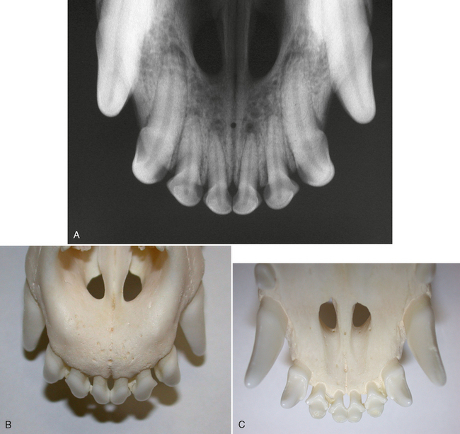
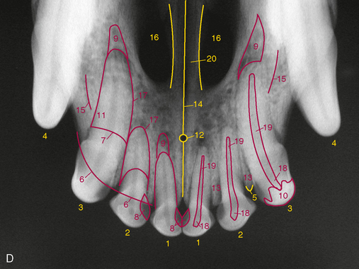
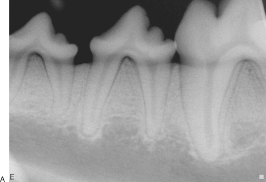
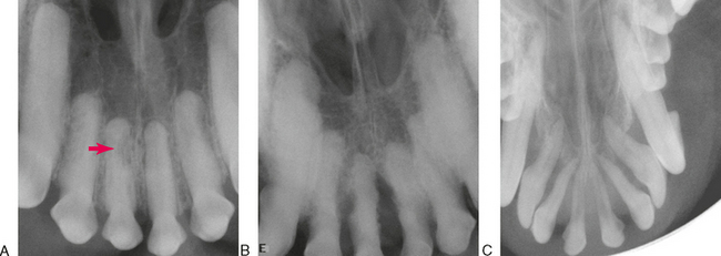
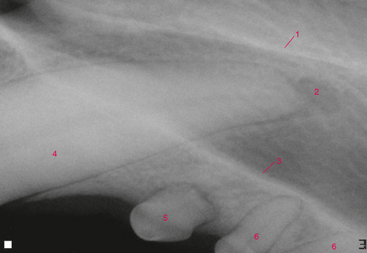
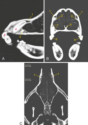
FIGURE 2-2 Normal incisor teeth. A, Radiograph of the incisor teeth and rostral maxillary region of a young adult dog. B, Dorsal view of prepared skull. C, Palatal (mirror) view of skull. D, Same radiograph as A. The crowns of the incisor teeth are foreshortened due to a projection angle that makes an image of the roots without elongation artifact (see Chapter 12).
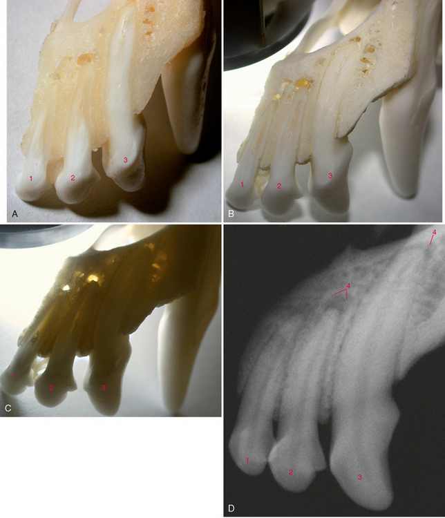
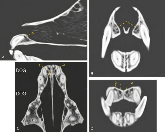
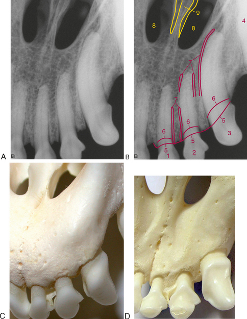
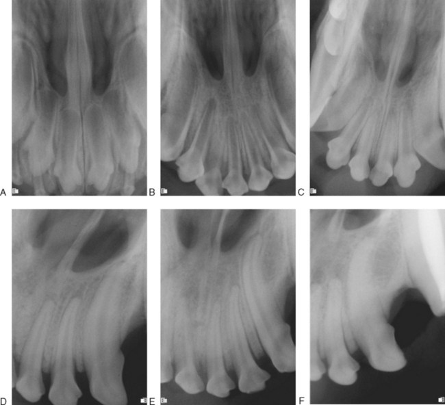
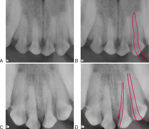
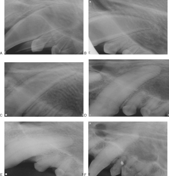
FIGURE 2-4 Effect of aging on the size of the pulp chambers and root canals of incisor teeth As teeth mature, secondary dentin is produced on the periphery of the pulp resulting in a progressively smaller pulp cavity with age. When the pulp experiences inflammation, dentin production can be accelerated resulting in a smaller pulp space compared to a healthy tooth. This can occur focally at the site of localized pulpitis. When it affects the entire pulp, the pulp appears more mature than normal. When a tooth pulp dies, maturation stops, making it appear less mature than normal (see Chapter 6). A, Three-month-old puppy. B, Five-month-old puppy. C, Eight-month-old puppy. D, Fifteen-month-old dog. E, Eighteen-month-old dog. F, Seven-year-old dog.
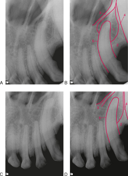
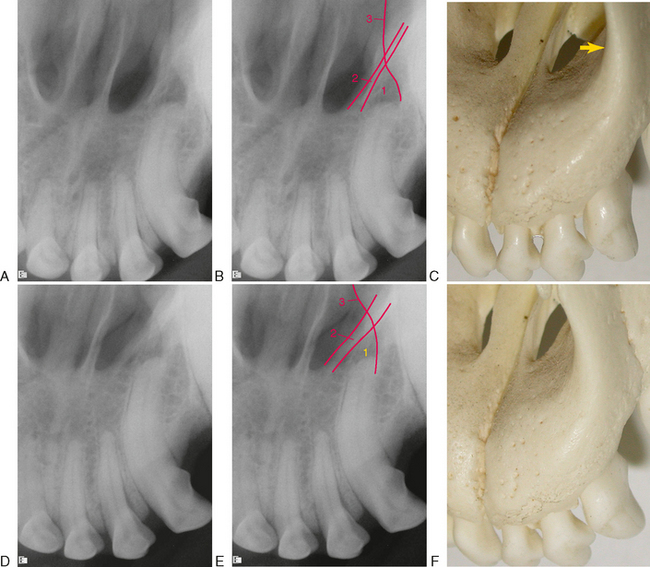
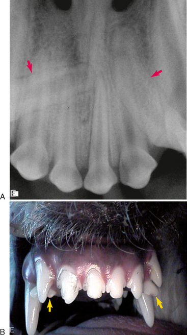
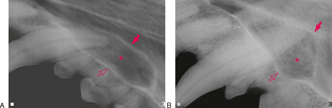
FIGURE 2-8 Another example of summation effect mimicking a periapical lucency A and B, Apparent radiolucency around the apex of the third incisor tooth. C, Skull showing how the nasal process of the incisive bone (arrow) makes a curved opacity that can appear to be the border of an endodontic lesion. D and E, The x-ray tube has been shifted to a more lateral position. The image of the incisive bone has shifted relative to the root apex, demonstrating that it is in a different plane from the root apex. The line of the incisive bone moved medially as the tube was shifted laterally indicating that the structure responsible for the linear opacity is labial to the tooth root (see Chapter 12). F, Skull showing how shifting the tube laterally moves the nasal process medially.
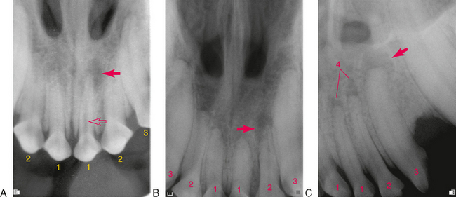
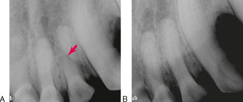
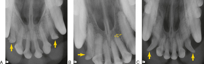
FIGURE 2-9 True lesions of endodontic origin A, Periapical lucency of the bone around the apex of the left upper first incisor tooth (arrow). Note also the relative immature (wider) root canal and pulp chamber compared to the other incisor teeth (open arrow), consistent with pulp necrosis and arrested dentin production. B, Periapical lucency of the bone around the apex of the left upper second incisor tooth (arrow). C, Periapical lucency of the bone around the apex of the left upper third incisor tooth (arrow). Note the more circular shape compared to the chevron lucencies on the first and second incisor teeth. The radiopaque border is consistent with a chronic endodontic lesion (see Chapter 6).
CANINE TEETH
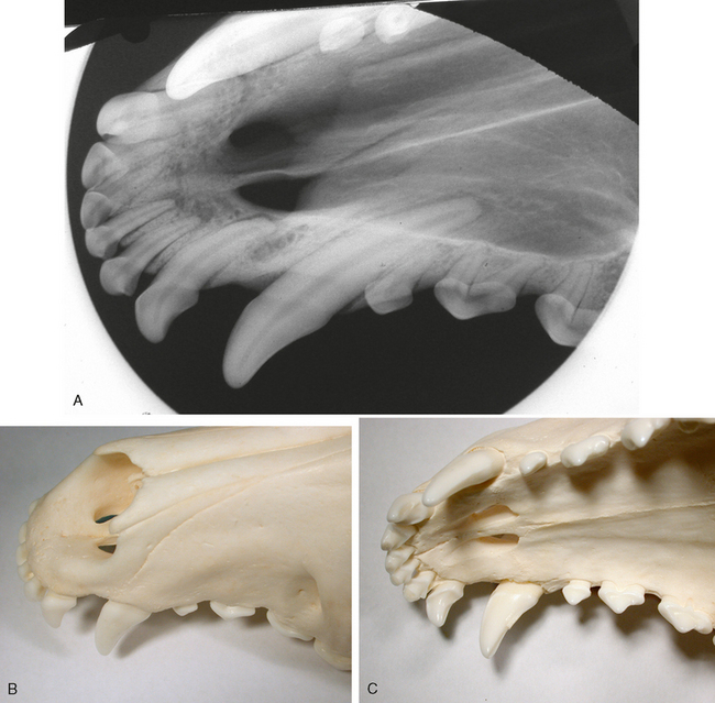
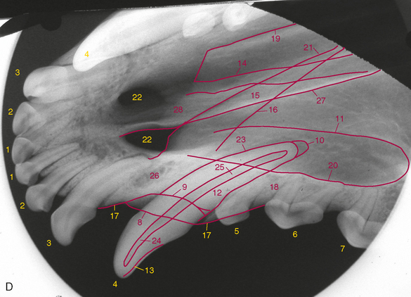
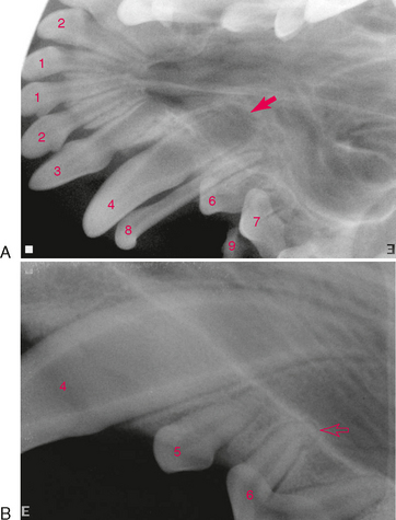
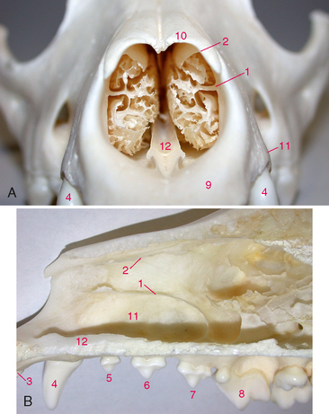
FIGURE 2-15 Normal dog canine tooth A, Radiograph of the skull of a young dog showing the canine tooth and surrounding structures. B, Dorsal view of prepared skull. C, Ventral (mirror) view of skull. D, Same radiograph as A.
MAXILLARY PREMOLAR TEETH
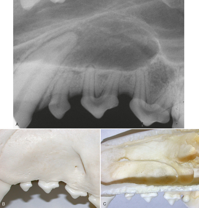
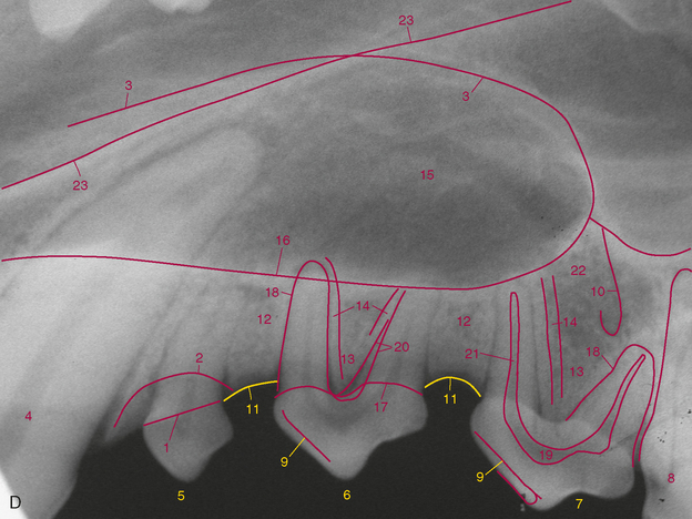
FIGURE 2-23 Normal maxillary premolar teeth The facial and palatal region of the dog consists of 36 bones designed to provide a large surface area for the sense of smell and to hold the teeth. The premolar teeth are all within the alveolar process of the maxilla. However, radiographs of the premolar teeth may project through the nasal, frontal, palatine, and zygomatic bones. A, Radiograph of the left maxillary premolar region of a young dog. B, Buccal (vestibular) view of prepared skull. C, Nasal surface of maxilla. D, Same radiograph as A. Correct use of the bisecting angle technique makes an image with accurate root length. The apical anatomy, however, will be slightly enlarged due to an increased object-to-film distance at the apex.
Stay updated, free articles. Join our Telegram channel

Full access? Get Clinical Tree


