CHAPTER 8 Swelling and Neoplasia
Some common causes of oral swelling include
Infection and Inflammation
Infections can result in oral swelling caused by inflammation or fluid accumulation. Infected root or bone fragments that persist after a tooth is lost are a common cause of swelling. Penetrating wounds or foreign bodies can also introduce bacteria into the deep oral tissues. Periodontal infections usually have adequate sulcular drainage to prevent swelling, but a periodontal abscess can occur if this outlet is blocked. Even with adequate drainage, a localized gingival swelling can occur in the form of a pyogenic granuloma. Focal fibrous hyperplasia can become large enough to mimic gingival tumors (Figure 8-1). Many of the lesions classified as fibromatous epulides are very likely fibrous hyperplasia. Endodontic infections can also present as swollen tissues if they are unable to establish drainage along the root or through a fistulous tract. A small localized swelling (parulis) can develop where a draining tract exits or is about to break open. When infection spreads from the initial site through the marrow spaces, it establishes osteomyelitis. This can manifest as an oral swelling (Figure 8-2) but can also occur with minimal or no tissue enlargement (see Chapter 11).
Oral and Dental Cysts
Apical radicular cysts (also called apical periodontal cysts, periapical cysts, and radicular cysts) are related to pulp inflammation or necrosis and are discussed in Chapter 6.
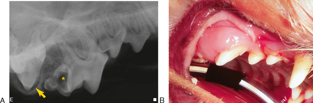
Lateral radicular cysts (sometimes called lateral periodontal cysts) are a type of odontogenic cyst that originates either from remnants of the Hertzwig epithelial root sheath (epithelial rests of Malassez) along the side of a root or possibly from remnants of dental lamina, which would make them similar in origin to adult gingival cysts (Figure 8-3). They are asymptomatic and often discovered late after they have attained a significant size, expanding the bone both labially and toward the nasal cavity (Figure 8-4). The most commonly identified lateral radicular cysts are associated with the maxillary canine teeth of dogs. Most are unilocular radiolucent lesions, but rarely there is a polycystic variant. Surgical debridement of the cyst lining with retention of the affected tooth is curative.
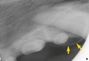
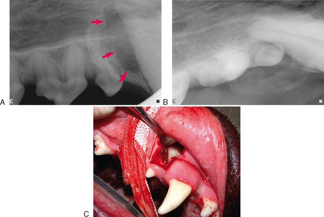
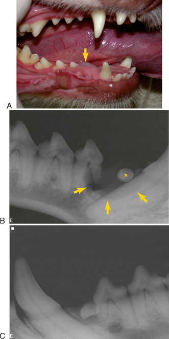
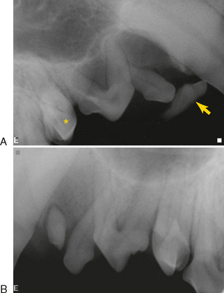
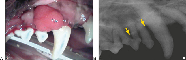
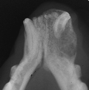
The dentigerous cyst is one type of odontogenic cyst called a follicular cyst that is an epithelium-lined sac surrounding the crown of an unerupted tooth. The epithelium is attached at the cervical area of the tooth. An eruption cyst (eruption hematoma) is a dentigerous cyst that is close to the surface surrounding a tooth that is eventually able to erupt, either naturally or with a minor operculectomy surgery or removal of the roof of the cyst (Figure 8-5). The tissue covering the teeth sometimes includes bone, in which case the redundant gingiva as well as the bone should be surgically removed to facilitate further tooth eruption. Deeper dentigerous cysts that surround an impacted tooth or one in which eruption has been irreversibly interrupted are relatively common, particularly associated with mandibular first premolar teeth (Figure 8-6). It can also occur with maxillary first premolar teeth and occasionally other teeth as well. Unerupted supernumerary first premolars can cause a cyst under the surface in an area where there is no tooth missing from the erupted dentition (Figure 8-7). Radiographs are not diagnostic for dentigerous cysts because odontogenic keratocysts, unilocular ameloblastomas, and other lesions may appear similar on radiographs. Dentigerous cysts often remain undiagnosed for many years, allowing them to become quite large, dissecting along the mandible, where they can interfere with the development and cause pressure resorption of the roots of other teeth in the quadrant (Figure 8-8). In early lesions, the crown often projects into the defect. Later, as the cyst changes location, the crown may no longer be within the lucency. Brachycephalic dog breeds seem to be predisposed to this, although they can be found in any breed. Many unerupted first premolar teeth remain quiet throughout life and do not develop dentigerous cysts (Figure 8-9).