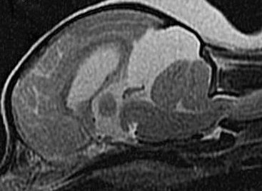
Intracranial Arachnoid Cysts in Dogs

Chapter 226


Intracranial Arachnoid Cysts in Dogs


Intracranial arachnoid cyst (IAC), also termed intraarachnoid cyst and quadrigeminal cyst, is a developmental brain disorder in which cerebrospinal fluid (CSF) is believed to accumulate within a split of the arachnoid membrane during embryogenesis. The developing neural tube is surrounded by a loose layer of mesenchymal tissue called the perimedullary mesh; this tissue eventually becomes the pia and arachnoid layers of the meninges. In normal development, pulsatile CSF flow from the choroid plexuses is thought to divide the perimedullary mesh into pia and arachnoid layers, effectively creating the subarachnoid space. It is postulated that some aberration of CSF flow from the choroid plexuses during this stage of development forces a separation within the forming arachnoid layer that eventually leads to the creation of an IAC. The intraarachnoid location of IACs has been demonstrated via light and electron microscopy in people.


The mechanisms by which an IAC continues to expand with fluid are unknown, but several theories have been proposed. Fluid may be secreted by the arachnoid cells lining the cyst cavity. There is evidence that cells lining the IAC may have secretory capacity. There also may be fluid movement into the cyst via an osmotic pressure gradient. Considering that the fluid within the IAC is nearly identical to CSF, this latter theory is unlikely. In addition, there have been documented cases in people in which the IAC and the subarachnoid space communicate via small slits. These slits act as one-way valves, diverting CSF into the cyst during systole but preventing return to the subarachnoid space during diastole.


Although IAC has been reported to occur in several locations in humans, all reported canine cases have been in the caudal fossa. Because IAC typically is associated with the quadrigeminal cistern in dogs, these accumulations of fluid often are called quadrigeminal cysts in this species. Also termed intracranial intraarachnoid cyst, IAC accounts for 1% of all intracranial masses in people and has been sporadically reported in dogs. IAC often is an incidental finding in humans; it has been suggested recently that this also may be the case in dogs.


Clinical Findings


In the veterinary literature, there have been nine clinical reports of IAC in dogs, with a total of 53 cases described. The IAC was suspected to be an incidental finding in approximately one third to over one half of the reported cases. The likelihood of clinical signs probably relates to the extent to which the cyst compresses the occipital lobe. The vast majority of reported IAC cases in dogs have involved small breeds, with a predominance of brachycephalic dogs. The breeds and numbers reported to date include the Shih Tzu (12), Maltese (4), pug (4), cavalier King Charles spaniel (4), Yorkshire terrier (4), Lhasa apso (4), Chihuahua (3), Staffordshire bull terrier (3), bulldog (3), Pekingese (2), West Highland white terrier (2), and one each of bichon frise, Pomeranian, cairn terrier, Jack Russell terrier, terrier mix, beagle, miniature schnauzer, and German shorthaired pointer.


There is a wide range in age at the time of clinical presentation (2 months to 10 years), with an approximate average age of 4 years. The most common clinical signs are attributed to forebrain dysfunction (including seizure activity), central vestibular (cerebellovestibular) disease, or both. Dogs also may be brought in with a primary complaint of neck pain.


Diagnosis of IAC typically is made via computed tomography or (preferably) magnetic resonance imaging (Figure 226-1). IACs also can be visualized using ultrasound imaging (via the foramen magnum, a temporal window, or a persistent bregmatic fontanelle), especially in younger dogs. The characteristic appearance of IAC is that of a large, well-demarcated fluid-filled structure that is isointense with the CSF spaces and is located between the caudal cerebrum and rostral cerebellum. Because IAC may be an incidental finding, it is important to rule out concurrent inflammatory disease (i.e., by CSF examination). In the author’s opinion, it is often difficult or impossible to discern whether IAC in the presence of another brain disorder is purely an incidental finding. Since the presence of a large fluid-filled structure within the cranial vault likely decreases intracranial compliance, some IACs may contribute to clinical signs rather than being simply an incidental finding. Because this disorder is believed to represent a developmental abnormality of the intracranial ventricular CSF system, it may occur concurrently with other fluid abnormalities (including congenital hydrocephalus). The cyst may or may not communicate with the remainder of the ventricular system. When one is faced with evidence of IAC and another disease (e.g., granulomatous meningoencephalitis) in the same patient, obtaining an optimal response to treatment may entail treating both conditions.


< div class='tao-gold-member'>

Stay updated, free articles. Join our Telegram channel

Jul 18, 2016 | Posted by in PHARMACOLOGY, TOXICOLOGY & THERAPEUTICS | Comments Off on Intracranial Arachnoid Cysts in Dogs

Full access? Get Clinical Tree

Get Clinical Tree app for offline access