CHAPTER 166 Inguinal Hernia
The extra-abdominal position of the testicles in horses necessitates a pathway for migration of the testes from their initial position in the abdomen to the scrotum. The testicle is encased in the vaginal tunic, which is the extra-abdominal continuation of the parietal peritoneum. The vaginal tunic traverses the inguinal canal, which is an obliquely oriented muscle plane between the external and internal abdominal oblique muscles. The inguinal canal is bounded by the superficial and deep inguinal rings, and entry into the inguinal canal is via a slitlike opening of the peritoneum, termed the vaginal ring. The size of the vaginal ring varies between breeds of horses and between individual horses of the same breed, but typically it is large enough to permit entry only of the normal testicular structures. Occasionally the vaginal ring is large enough to permit herniation of small intestine into the vaginal tunic, wherein the intestine descends alongside the spermatic cord toward the testicle in the scrotum. The size of the vaginal ring relative to the anatomic structures that traverse it determines whether a hernia is strangulating or nonstrangulating. This type of hernia is termed an indirect hernia, nomenclature that is borrowed from human terminology. A direct hernia occurs when the fascia adjacent to the vaginal ring ruptures and herniated bowel enters the inguinal canal; however, this bowel is not contained within the vaginal tunic. In horses, inguinal hernias are almost exclusively indirect hernias. In common usage, the terms scrotal hernia and inguinal hernia are used interchangeably. Inguinal hernias in horses may be either congenital or acquired. The signalment, clinical signs, and treatment differ for congenital and acquired inguinal hernias and will be discussed separately.
CONGENITAL INGUINAL HERNIA
Diagnosis
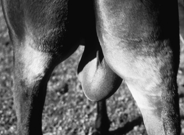
Congenital inguinal hernias are usually evident at birth in male horses. The condition arises because a larger-than-normal vaginal ring allows small intestine to exit the abdomen, pass through the superficial and deep inguinal rings, and descend into the scrotum. The bowel is contained inside the parietal vaginal tunic. Congenital hernias are usually nonstrangulating because the vaginal ring is larger than normal and the size of the vaginal tunic limits the length of bowel that can herniate. Anecdotal evidence suggests that Standardbreds and draft breeds have a much higher prevalence of congenital inguinal herniation than other breeds. Affected horses may have unilateral or bilateral involvement. The diagnosis is made by external palpation of the enlarged scrotum (Figure 166-1). The testicle can be palpated within the scrotum along with one or several loops of flaccid small intestine. The contents of the hernia should be easily reducible by external massage of the scrotum. Reduction of the hernia may be facilitated by positioning the foal in dorsal recumbency. Foals with uncomplicated inguinal herniation should have no clinical signs other than scrotal enlargement.
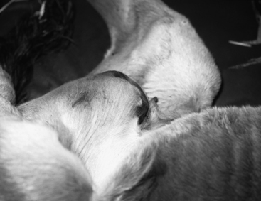
Occasionally the vaginal tunic may rupture in foals with inguinal hernias. Small intestine that would normally be confined within the vaginal tunic is able to exit the vaginal tunic via this rent and enter the subcutaneous space in the scrotum, inguinal area, and medial aspect of the thigh. In this situation, the hernia is no longer reducible, and skin in the inguinal area becomes erythematous and bruised (Figure 166-2). The vascular supply to the bowel becomes strangulated, and the foal develops signs of colic. Careful observation will reveal that the enlargement that accompanies the hernia is no longer confined to the scrotum, as the jejunum dissects cranially in the subcutaneous space. Peristalsis of the jejunum can be observed through the inguinal skin. Rents in the vaginal tunic usually occur in neonates and, rarely, in foals older than 1 week. Once identified, this condition is an emergency and warrants immediate surgical correction.
Treatment
Conservative Treatment
If the hernia is reducible, initial management of a foal with a nonstrangulating congenital hernia is nonsurgical and consists of frequent manual reduction of the herniated intestinal contents, which is facilitated by placing the foal in dorsal recumbency. Foals with hernias that are small or intermittent are very likely to spontaneously resolve. Studies to document the success rate for spontaneous resolution of congenital inguinal hernias are not available to my knowledge; however, clinical experience suggests that in most affected foals the condition resolves without surgical intervention within the first weeks to months of life. It is unclear whether the hernia resolves because of manual reduction or despite it. Frequent daily attempts at reduction do force the owner to watch the foal more closely.
Stay updated, free articles. Join our Telegram channel

Full access? Get Clinical Tree


