Infrequently Encountered Gram-Negative Rods

Chapter 27 Infrequently Encountered Gram-Negative Rods


BARTONELLA SPECIES


Bartonella is the only genus of the family Bartonellaceae. These aerobic, slowly growing, facultatively intracellular, gram-negative organisms are placed in the α-division of the Proteobacteria based on their 16S rDNA sequences. The genus includes bacteria formerly classified in the genera Grahamella and Rochalimaea; some of the 18 species are presented in Table 27-1. All are fastidious, argyrophilic, hemotrophic, and highly adapted to a mammalian reservoir host. They cause a variety of clinical syndromes in both immunocompetent and immunocompromised humans and animals. Diseases caused by bartonellae are usually vector-borne, with transmission by arthropods (lice, fleas, ticks, biting flies, mites, chiggers). Transmission as a result of blood transfusions has also been documented.


Bartonella bacilliformis is the agent of classic human bartonellosis. This disease occurs in regions of South America and manifests as progressive anemia (Oroya fever) or as cutaneous disease (verruca peruana).


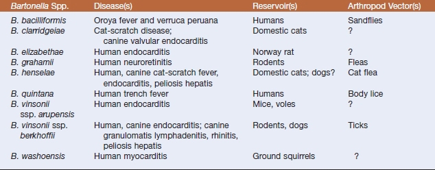
Bartonella henselae is the etiologic agent of cat-scratch fever, which is also transmitted by cat fleas. Manifestations of human and canine disease may include prolonged fever, meningitis, bacillary angiomatosis, and peliosis hepatis. Bacillary angiomatosis is a neovascular proliferative disorder that may involve skin, regional lymph nodes, liver, spleen, brain, bones, bowel, and lungs. Peliosis hepatis–affected tissue contains numerous blood-filled partially endothelial cell-lined cystic structures. Chronically bacteremic cats are the reservoir. Bartonella henselae may also be an important emerging pathogen of dogs. Polyarthritis, weight loss, seizures, epistaxis, and endocarditis may result from inoculation by way of cat scratches.


Bartonella quintana causes human trench fever, which was a leading cause of morbidity among Allied troops in World War I. Lymphadenopathy and bacillary angiomatosis have also been related to this agent, which is transmitted by the human body louse.


Based on current evidence, Bartonella vinsonii ssp. berkhoffii is the species most often associated with canine disease, which manifests as endocarditis, myocarditis, and granulomatous lymphadenitis. Endocarditis, typically preceded by fever of unknown origin and intermittent leg lameness, occurs mainly in large-breed dogs, and there is a predisposition to aortic valve disease. Focal areas of myocardial inflammation can be found in dogs infected with B. vinsonii ssp. berkhoffii. This organism has also been recovered from a human endocarditis patient.


Both Bartonella clarridgeiae and B. henselae can be maintained as intravascular microflora of apparently healthy cats for years. Persistent infection in cats may also yield chronic disease manifestations. There may be an association between high antibody titers and increased risk for development of renal and urinary tract abnormalities. Chronic Bartonella infection may also be a cause of uveitis and myocardial inflammation.


Pathogenesis of Bartonella-induced disease in animals remains to be elucidated. It likely involves bacterial dissemination from regional lymph nodes and the bloodstream, or via the portal system following enteric infection, but virulence determinants have not been clearly defined. Bartonellae interact with both erythrocytes and endothelial cells. Mechanisms of deformation, invasion, and proliferation have been studied, and an extracellular protein, deformin, in B. bacilliformis, produces deep invaginations of the red blood cell membrane. These invaginations are likely portals of entry for invading bacteria. Polar flagella have been implicated in adhesion and erythrocyte invasion. Bundle-forming pili have been described in B. henselae and B. quintana, and appear to play an important role in host cell attachment. Bartonella henselae undergoes phase variation in its outer-membrane proteins, which may facilitate immune evasion. This organism is unable to synthesize heme, but possesses a protein that is involved in heme acquisition. Induction of endothelial cell proliferation is common to many pathogenic bartonellae. Colonization and inva sion of vascular endothelium is a crucial step in the establishment of proliferative lesions by B. bacilliformis, B. henselae, and B. quintana. Proliferation of nonlymphoid cells results in formation of new blood vessels, and this angioproliferation might be a strategy for expanding bacterial habitat in vivo.


Bartonella quintana appears to be able to inhibit apoptosis, which may enhance its intracellular survival and proliferation. Its intraerythrocytic localization is a unique strategy for bacterial persistence. Colonization of red blood cells in the absence of hemolysis preserves the bacteria for efficient vector transmission, provides protection from the host immune response, and possibly contributes to the decreased efficacy of antimicrobials in eradicating the infection.


Diagnosis of bartonellosis may be extremely difficult because the typical clinical features are not distinctive. Etiologic diagnosis is usually established by serologic testing and detection of the organisms by bacteriologic culture, polymerase chain reaction (PCR) assays, and histologically with Warthin-Starry silver staining or immunohistochemistry. Serologic testing is by an immuno-fluorescence assay, in which cross-reactions with Coxiella burnetii and chlamydiae may occur. Diagnosis relies heavily on the detection of the agent or its DNA in affected tissue.


Lymph node biopsies or aspirates, affected portions of liver or spleen, and blood are the specimens of choice. Efficiency of blood culture is increased by lysis of erythrocytes. Blood collected in EDTA is held at −70° C for 24 hours before inoculation onto plated isolation media. Colonies of Bartonella spp. appear in 10 days or more on solid media, and freshly prepared heart infusion agar with 5% to 10% horse or rabbit blood supports better growth than chocolate or sheep blood agar. Primary culture plates should be sealed against dehydration after 24 hours’ incubation and incubated for up to 6 weeks before being discarded as negative. Growth is optimal at 37° C in 5% to 7% CO2 for all species except B. bacilliformis, which has an optimal growth temperature of 25° to 28° C and grows best without supplemental CO2.


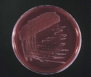
Bartonella colonies usually exhibit two morphotypes (Figure 27-1). One is irregular, whitish, raised, rough, and dry, and may be cauliflower-like in appearance. The other type is small, tan, circular, moist, entire, and adherent to the agar. Gram stains of the colonies reveal tiny, slightly curved, faintly staining, gram-negative bacilli. Most Bartonella spp. are oxidase, catalase, urease, and nitrate reductase negative, and carbohydrates are not utilized. Genus and species iden tification is best accomplished by molecular methods.


Bartonellae are highly susceptible in vitro to some β-lactams, aminoglycosides, macrolides, and tetracyclines, but less so to penicillin and clindamycin. Considerable variation occurs in susceptibility to fluoroquinolones. Azithromycin is probably the drug of choice for treatment of canine bartonellosis. Evidence suggests that Bartonella spp. may be transmitted by fleas and ticks to cats and dogs, so use of insecticides and acaricides may be important in prevention and control.


CDC GROUP EF-4A


A group of gram-negative, nonmotile, coccoid to short, facultatively anaerobic bacilli has been assigned by the Centers for Disease Control and Prevention (CDC) to a group designated eugonic fermenter (EF)-4A. These organisms share phenotypic features with Pasteurellaceae, but are members of the emended family Neisseriaceae. Group EF-4A bacteria are part of the normal flora of the oral and nasal cavity of cats, dogs, and rodents. However, they have been recovered from bite wounds and scratches in man and animals and are associated with acute pneumonia of domestic cats and dogs and captive felids. Pulmonary disease is rather distinctive, in that lesions are multifocal, granulomatous, and involve all lung lobes. There are also rare extrapulmonary infections such as purulent peritonitis, hepatic abscessation, retrobulbar abscesses, otitis, and sinusitis.


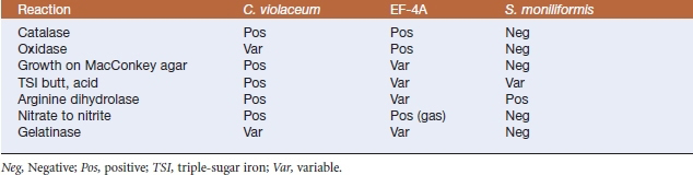
Diagnosis is by culture of affected tissues. After 48 hours of aerobic incubation on blood agar plates, colonies are 1 mm in diameter, nonhemolytic, convex, round, opaque, and nonpigmented to yellowish with a distinctive popcornlike odor. Isolates are catalase and oxidase positive, and growth is sometimes seen on MacConkey agar. Triple-sugar iron (TSI) agar (butt and slant) have an initial weak acid reaction. Other distinguishing features include nitrate reductase, gelatinase, and arginine dihydrolase activities, acid production from glucose, and inability to produce urease or indole (Table 27-2).


These organisms are reported to be susceptible in vitro to ampicillin, aminoglycosides, sulfa drugs, and tetracyclines. Resistance to narrow-spectrum cephalosporins, trimethoprim-sulfamethoxazole, penicillin, and erythromycin has been reported.


THE GENUS CHROMOBACTERIUM


Violet-pigmented bacteria, described since the late 1800s as a cause of fatal septicemias in animals and humans, have now been placed in the genus Chromobacterium, which is the closest neighbor to the Neisseriaceae. Chromobacterium violaceum, the sole species in the genus, is a motile, facultatively anaerobic, short to medium-length, gram-negative rod. Pigmentation is caused by the production of a nondiffusible pigment, violacein; nonpigmented strains are quite rare.


Chromobacterium violaceum is a saprophyte inhabiting soil and water, but is also an opportunistic pathogen of extreme virulence, especially in compromised hosts. Sporadic cases have been reported worldwide, primarily from tropical and subtropical regions. Infections in swine, dogs, buffaloes, Barbary sheep, nonhuman primates, and a panda have resulted in necrotizing pleuropneumonia, septicemia, and skin, pulmonary, splenic, or hepatic abscesses. Most human infections are trauma associated, with skin as the portal of entry, but infection may also be by the oral route.


Little is known about pathogenesis. The organism has elastase activity in vitro, which may in part explain its ability to produce necrotizing lung lesions. Violacein is cytotoxic in vitro, especially for fibroblasts, which are primary components of the integument.


Diagnosis is based on bacterial isolation and identification. Colonies are 0.5 to 1.5 mm in diameter, smooth, and convex with entire edges and β-hemolysis. Growth may occur at 25° and 42° C. A faint violet tint generally develops after 24 hours’ incubation on blood agar, and the color becomes deeper in subsequent days. Colonies on MacConkey agar are colorless. Most isolates produce oxidase and catalase, but are negative for Voges-Proskauer reaction and do not hydrolyze esculin. There is an acid reaction on TSI slants, but H2S is not produced. Arginine dihydrolase is produced. Oxidase-positive nonpigmented strains can be differentiated from Aeromonas or Vibrio spp. by the ability of the former to grow in nutrient broth without salt and to ferment glucose, mannitol, and maltose, and by their lack of lysine and ornithine decarboxylase activities. Oxidase-negative strains can be mistaken for enteric bacilli (see Table 27-2).


Chromobacterium violaceum is susceptible to fluoroquinolones and tetracyclines, variably susceptible to aminoglycosides, but commonly resistant to β-lactam antibiotics.

< div class='tao-gold-member'>

Stay updated, free articles. Join our Telegram channel

Jul 18, 2016 | Posted by in PHARMACOLOGY, TOXICOLOGY & THERAPEUTICS | Comments Off on Infrequently Encountered Gram-Negative Rods

Full access? Get Clinical Tree

Get Clinical Tree app for offline access