Fig. 25.1
Light micrographs of mouse kidneys prepared with the “in vivo cryotechnique ” (IVCT) or resection of fresh tissues and quick-freezing (RF-QF), followed by FS. (a, b) Serial sections are stained with hematoxylin-eosin (a, HE) and immunostained for phospho-(Ser/Thr) protein kinase A/C substrate (b, P-PK-S). The P-PK-S-immunopositive areas are mostly observed in the cortex (Cx). Inset of (b) is the immunocontrol for P-PK-S in the renal cortex at the same magnification of (b). (c–h) Normal kidneys on serial sections stained with HE and immunostained for P-PK-S, by two different procedures with IVCT-FS and RF-QF-FS. (c–e) With IVCT-FS, immunolocalization for P-PK-S is detected in the cytoplasm of the proximal tubule s (d and e, arrows), and each tubular cell shows different intensities of immunostaining. (e) Another higher magnification view shows heterogeneous immunolocalization patterns of P-PK-S in the cytoplasm. (f–h) With RF-QF-FS, the immunolocalization of P-PK-S is detected along the basal striations of the renal proximal tubule s (g and h, arrows). Cx cortex, G glomerulus . Bars 300 m (a, b), 100 m (c, d, f, g), 50 m (e, h)
25.3 Immunolocalization of P-PK-S in Hypoxic Mouse Kidneys Prepared with IVCT-FS
To examine the transient effects of hypoxia on the immunolocalization of P-PK-S, both the morphology of HE-stained tissue and the immunolocalization of P-PK-S, NBC1 (an ion cotransporter), and 4.1B (a membrane skeletal protein ) (data not shown) were compared in serial sections from renal proximal tubule s under various hypoxic condition s (Fig. 25.2). Under normal blood circulation , P-PK-S was immunolocalized diffusely throughout the cytoplasm of most epithelial cell s in the proximal tubule s and occasionally along the basal striations (Fig. 25.2b, d). The diffuse immunolocalization of P-PK-S in the proximal tubules as shown in Fig. 25.2b, d was very similar to that shown in Fig. 25.1e. After 10 or 30 s hypoxia, their signals were also detected in the proximal tubules, some of which were immunolabeled with anti-NBC1 or 4.1B antibody , showing the proximal S1–S2 segment s . Higher magnification demonstrated that immunolocalization of P-PK-S in the cytoplasm was almost identical to that under normal blood circulation (data not shown). After 1 or 2 min hypoxia, it was also detected in the proximal tubules at low magnification (Fig. 25.2g, h). Higher magnification showed that it had changed to become localized, mainly in the basal striations (Fig. 25.2i, j). In contrast, the immunolocalization of both NBC1 and 4.1B was always detected in the basal striations of the S1–S2 proximal tubules under all conditions (NBC1: Fig. 25.2e, j, o). These findings indicate that immunolocalization of P-PK-S was almost completely changed from the whole cytoplasm to the basal striations in the proximal tubules after 1 min hypoxia.
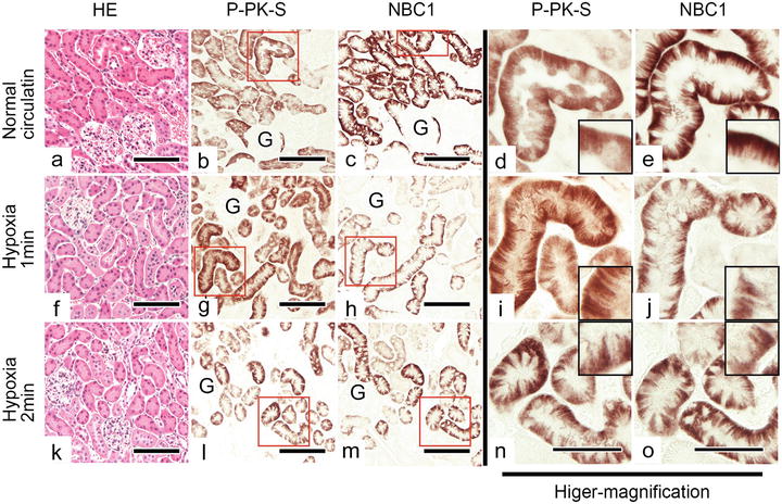
Fig. 25.2


Light micrographs of mouse kidney tissue specimens prepared with IVCT-FS under normal blood circulation (a–e) and hypoxia of 1 min (f–j) and 2 min (k–o) before the cryofixation . Serial sections are stained with HE (a, f, k) and immunostained for P-PK-S (b, d, g, i, l, n) and Na+/HCO3 − cotransporter (NBC)1 (c, e, h, j, m, o). The tissue areas of P-PK-S-immunopositive proximal tubule s are more widely distributed than those of NBC1 . Insets show the basal cytoplasm of the epithelial cell s , thus indicating the immunolocalization of the P-PK-S in the whole cytoplasm (b, d) under normal blood circulation along the basal striations (g, i, l, n) under 1 and 2 min hypoxia. The NBC1 is almost always immunolocalized along the basal striations (c, e, h, j, m, o). G glomerulus . Bars 100 μm (a–c, f–h, k–m), 50 μm (d–e, i–j, n–o)
Stay updated, free articles. Join our Telegram channel
Full access? Get Clinical Tree
