Imaging in Diagnosis of Endocrine Disorders

Chapter 40


Imaging in Diagnosis of Endocrine Disorders


Imaging Modalities in Endocrinology


Radiography has little value for imaging endocrine organs and is mainly used to detect lung metastases. Ultrasonography (US) is the most commonly used imaging modality for diagnosis of endocrine disorders because of its availability and relative low cost. Major endocrine organs such as the thyroid gland, parathyroid glands, and adrenal glands are superficially located, which makes them easily accessible with US using high-resolution transducers (≥10 MHz). Additionally, US allows for fine-needle aspiration, tissue-core biopsy, or interventional procedures of endocrine organs to be performed in real-time. Two newer US techniques, which are still under clinical investigation, can help to improve the accuracy of gray-scale diagnostic US. Contrast-enhanced US involves intravenous injection of gas microbubbles as vascular contrast agents to improve the detection of perfusion and vascularity of both normal and abnormal organs. Changes in vascularity and blood flow are frequently seen secondary to pathology and are represented in time-intensity curves as alterations of the shape of the curve on contrast-enhanced US. The second method, elastography, is an US technique that evaluates the stiffness of tissues by measuring the displacement of ultrasound echoes before and after compression. Elastography thereby provides information about the mechanical properties of tissues and is currently used in people to differentiate malignant from benign lesions in thyroid tissue.


Computed tomography (CT) and magnetic resonance imaging (MRI) are very useful for evaluation of the extent, local invasiveness, and local or distant metastases of neoplastic processes. CT is the modality of choice for detection of lung metastases, whereas MRI provides excellent delineation of anatomic structures because of inherent high-contrast resolution and is the modality of choice for evaluation of presurgical tissue and vascular invasion. Both modalities allow visualization of intracranial structures and are particularly suited for imaging the pituitary gland. Dynamic CT contrast studies including targeted angiography or dual-phase CT are useful for examination of adrenal glands, pituitary gland, and pancreatic disorders.


The routine use of nuclear medicine in veterinary endocrinology is limited to thyroid scintigraphy. This modality is extremely useful to determine unilateral or bilateral lobe involvement, the status of the gland, the location of hyperfunctioning ectopic or accessory tissue, and distant metastases of thyroid tumors. Availability of scintigraphy is limited in veterinary medicine. Box 40-1 lists some useful imaging tips for evaluating endocrine disorders in dogs and cats.


Imaging the Thyroid Gland


The thyroid gland is composed of two separate lobes except in a few large-breed dogs in which an isthmus connects both lobes caudally. Thyroid lobes are oblong and span the dorsolateral aspect of the trachea, medial to the common carotid arteries, from the first to the eighth tracheal ring. The thyroid gland is supplied with arterial blood from a large cranial and a smaller caudal thyroid artery, both branches of the common carotid artery. The caudal thyroid artery is absent in cats. Venous drainage is via the cranial and caudal thyroid veins.


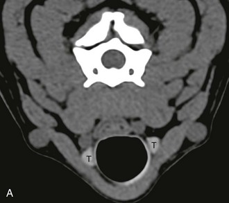
US is the primary modality for evaluation of the thyroid gland. The gland appears on US as a homogeneous, well-delineated, hyperechoic (compared with the surrounding musculature), fusiform structure. Measurements of the height (dorsoventral axis) of the thyroid gland and calculation of its volume using the formula of a rotation ellipse have the lowest variability and should be preferred in comparative and follow-up studies. On CT, the normal thyroid gland is homogeneous, ovoid or triangle-shaped, and hyperattenuating compared with the surrounding tissues (Table 40-1). The hyperattenuation is due to its natural high iodine content, which is an element with a high atomic number (53) compared with most other elements in the body (Figure 40-1, A). After intravenous injection of iodinated contrast medium, the gland shows usually diffuse enhancement. On MRI, the thyroid gland may appear heterogeneous, particularly on T2-weighted images, or homogeneous. On T1-weighted images, the thyroid gland is isointense to surrounding muscles in half of cases and shows intermediate signal intensity between fat and muscle in the other half. After contrast medium administration (gadolinium-based), the thyroid gland shows intensity between muscle and fat or is isointense to fat. On T2-weighted images, the thyroid gland shows intensity between muscle and fat. MRI is the best modality to evaluate local tissue invasion, detect cervical lymphadenopathy, and detect recurrent thyroid carcinoma after treatment.


Radionuclide iodine (123I, 131I) or pertechnetate (99mTcO4) is taken up by thyroid tissue and can be used for thyroid scintigraphy. In contrast to pertechnetate, iodine isotopes are incorporated in thyroglobulin (organification), enabling the determination of “true” uptake, which could be more reflective of thyroid physiology. Despite this advantage of iodine, pertechnetate routinely is preferred because it is easily obtainable from an in-house molybdenum generator, is cheaper, and has a shorter half-life compared with iodine isotopes. On a normal scintigraphic study, the thyroid-to-salivary uptake ratio is less than 1. Injection of iodinated contrast medium on CT scan influences the results of nuclear imaging for 6 to 8 weeks. Gadolinium-based MRI contrast agents do not interfere with thyroid function and subsequent nuclear imaging.


Canine Thyroid Neoplasia


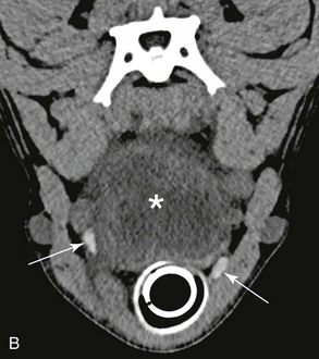
In dogs, thyroid neoplasia (mostly carcinomas, less commonly adenomas) is associated with a unilateral (66%) or bilateral (33%) mass caudal to the pharynx. Clinically detectable neoplasms usually are nonsecreting, resulting in euthyroidism throughout the course of the disease. In dogs with thyroid neoplasia, diagnostic imaging is used (1) to define the thyroid origin of the cervical mass, (2) to detect local or distant metastases, and (3) to evaluate local tissue invasion. Whatever imaging modality is used, it is sometimes difficult to document the thyroidal origin of the mass when its size severely disrupts the normal anatomy of the cervical area. Scintigraphy, CT, and to a lesser extent US may be indicated to determine whether large cervical masses arise from the thyroid gland or from other tissues. When the mass arises from tissues other than the thyroid, both thyroid lobes should be visible exhibiting a normal pattern (Figure 40-1, B). Additionally, US and CT can be used to guide a fine-needle aspiration or a core biopsy of a cervical mass.


With thyroid neoplasia, radiography may reveal a (sometimes mineralized) mass and possibly show displacement or compression of the trachea or the esophagus (mostly ventrally) or deformation of the larynx. Thyroid neoplasia appears as a large, heterogeneous mass involving most commonly the entire lobe or gland, with variable delineation, hypoechoic (US), isoattenuating to hypoattenuating (CT), or hyperintense (all routine MRI sequences); the mass sometimes may contain multiple cystic areas of necrosis or hemorrhage that alternate with areas of normal parenchyma, dense connective tissue, or mineralization. Thyroid carcinoma shows strong vascularization on power or color Doppler, contrast-enhanced US, and contrast-enhanced CT. CT is highly specific (100%) and MRI is highly sensitive (93%) in diagnosing thyroid carcinoma, whereas US has a moderate sensitivity (79%) and a poor specificity (33%). Main differential diagnosis is a carotid body tumor. On scintigraphy, thyroid tumors are of various sizes with irregular areas of pertechnetate uptake and usually heterogeneous distribution of radioactivity. Diffuse increased and decreased uptake patterns also have been described. If the tumor is secreting excessive amounts of thyroid hormones, moderate to extensive areas of increased, usually uniform, tracer uptake are detected, and the contralateral lobe exhibits suppressed uptake because of negative feedback onto the pituitary gland and resultant lack of thyroid-stimulating hormone (TSH) secretion. Unfortunately, increased uptake of radionuclide does not always correlate with increased production of thyroid hormones by the tumor. For instance, if the thyroid tumor destroys enough of the thyroid gland (≥75%) to cause subnormal thyroxine concentration, the pituitary gland increases its TSH release, and the remaining normal tissue is stimulated. The nonsecreting thyroid neoplasm shows decreased uptake, and the remaining normal tissue has increased uptake. Diagnostic imaging cannot reliably differentiate adenoma from carcinoma.


Distant pulmonary metastases from local invasion of the thyroid veins are common, and thoracic radiographs or lung CT scan should always be performed in cases of thyroid carcinoma. If a cranial mediastinal mass is present, neoplastic transformation of ectopic thyroid tissue should be considered. The second most common site of metastases is the retropharyngeal lymph nodes, which are best imaged on CT, MRI, and Doppler or contrast-enhanced US. Other sites of metastatic spread include abdominal organs (liver, kidneys, spleen, and adrenal glands), justifying standard abdominal US or CT to be performed, and also bone, bone marrow, brain, and spinal cord. Thyroid scintigraphy is a specific tool for identification of metastasis but is not considered sensitive. Scintigraphic visualization of metastases in the presence of an intact trapping mechanism in thyroid tumor cells indicates a high trapping ability of iodine in the tumor tissue, and this may be considered a predictive factor of radioiodine therapy effectiveness.


Even with the use of US, CT, or MRI, the detection of local tissue invasion by a thyroid carcinoma may be challenging. US is less sensitive than MRI and CT for detecting capsule disruption and local tissue invasion. US strongly depends on the skill of the operator and the quality of the US equipment, and it is limited for detection of retrotracheal and intrathoracic extension of the thyroid malignancy. The extension of tumor into adjacent soft tissues and vessels of the neck is the main purpose of CT and MRI in assessing patients with thyroid carcinoma, with MRI having the best contrast resolution. Posttreatment imaging should be performed at 3 to 6 months and include thoracic radiography and pertechnetate scintigraphy.

< div class='tao-gold-member'>

Stay updated, free articles. Join our Telegram channel

Jul 18, 2016 | Posted by in PHARMACOLOGY, TOXICOLOGY & THERAPEUTICS | Comments Off on Imaging in Diagnosis of Endocrine Disorders

Full access? Get Clinical Tree

Get Clinical Tree app for offline access