35 Iatrogenic tooth damage
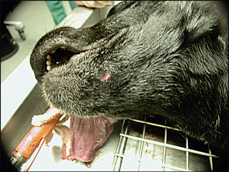
ORAL EXAMINATION – UNDER GENERAL ANAESTHETIC
In summary, examination under general anaesthesia identified the following:
RADIOGRAPHIC FINDINGS
The following were evident on the radiographs:
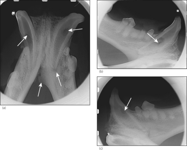
2. Teeth 304 and 404 were immature teeth with wide pulp system diameter and incomplete apical closure (Fig. 35.2a–c)

Figure 35.2 Radiographs of the rostral mandible.
(a) Rostrocaudal view. Although the dog is 6 years old, 304 and 404 are immature teeth (wide pulp system diameter and incomplete apical closure). The development of these teeth is compatible with a dog younger than a year of age. Consequently, pulpal inflammation and necrosis (which stops further tooth development) must have occurred at around 9 months of age. Note the circular lucencies at the midroot of both 304 and 404. These are compatible with drill holes for a pin or a screw to repair the rostral mandibular fracture. The pulpal inflammation and necrosis of 304 have spread to involve the periapical tissue, as evidenced radiographically by the radiolucent zone around the apex of the root. The periosteal new bone formation on the left is a consequence of the pulp and periapical disease of 304.
Stay updated, free articles. Join our Telegram channel

Full access? Get Clinical Tree


