22 Hypocalcaemia in a green iguana
Introduction
Green iguanas are common reptile pets (Box 22.1). Destined to become large (<2 m in length) powerful animals, many owners struggle to provide an appropriate environment for them as they mature. This may lead to husbandry deficiencies as in this case and potentially fatal disease.
BOX 22.1 Ecology of green iguanas
• Range in the wild covers Central and South America, and the Caribbean. Introduced feral populations exist in the USA in south Florida, Hawaii and the Rio Grande Valley of Texas
• Tail: undergoes autotomy; re-grown tail will contain cartilaginous vertebrae. Will swim, using lateral tail movement for propulsion. Use tail to ‘whip’, protecting from predators; also use teeth and claws for protection
• Normal habitat: arboreal species – they are agile climbers using their long claws for grip. Rubbing against rough surfaces also helps to remove old skin during ecdysis. Diurnal
• Free-ranging diet: herbivorous, feeding on leaves, flowers, fruit and growing shoots of many plant species. Occasionally consume invertebrates (usually as a by-product of eating plant material). Juveniles more likely to consume invertebrates
• Gender determination: sexual dimorphism. Males have well-developed dewlaps, used to regulate body temperature and for courtship and displays. Males also have prominent femoral pores on their ventral thighs. Hemipenal bulges present caudal to the cloaca in males. Adult males are highly territorial, particularly during the breeding season
• Reproduction: oviparous (egg-laying). Clutch of up to 40 eggs, laid in a nesting burrow. Temperature-dependent sex determination. Incubation 10–15 weeks. Young animals often remain in a group
Clinical Examination
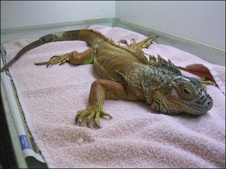
• Evidence of previous autotomy, with tail regrowth (obvious change to skin scales part-way down the tail, with smaller scales distal to the site of tail loss) (Fig. 22.1)
• Mandibular shortening and widening (likely associated with metabolic bone disease occurring as a growing individual)
• Unable to support body weight and limbs flaccid on presentation (Fig. 22.1). Paresis diagnosed (rather than ataxia)
Stay updated, free articles. Join our Telegram channel

Full access? Get Clinical Tree


