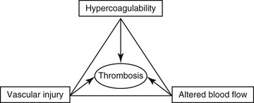
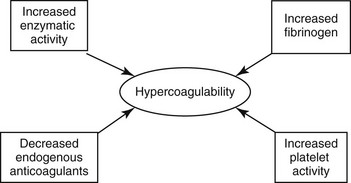
Hypercoagulable States

Chapter 64


Hypercoagulable States


A hypercoagulable state is an enhanced tendency to form venous or arterial thrombi. In 1845 a young German pathologist named Rudolf Virchow presented a novel and relatively simple concept of the pathogenesis of thromboembolic disease, which remains valid to this day. Virchow described three factors (Figure 64-1) that are the core reasons for the development of venous thrombosis: alterations of the blood (hypercoagulability), impairment of blood flow (stasis), and changes in the vessel wall (vascular injury). Research into the molecular basis of thrombosis has since enhanced our understanding of the pathophysiology of each of these concepts and thereby also our ability to diagnose and treat patients with thromboembolic disease. This chapter focuses primarily on the first of the three legs of Virchow’s triad—hypercoagulable states—but briefly describes the effects of stasis and disruption of the intact endothelium.


Pathophysiology


Impairment of normal blood flow (stasis) leads to accumulation of procoagulant factors and facilitates increased interaction between the platelets and their receptors on the vessel and increased exposure to procoagulant factors released by the endothelium, which promotes thrombus formation. If prolonged or severe, stasis may cause acidosis and tissue hypoxia, which lead to vascular injury. Stasis, along with hypercoagulability, plays an important role in the development of venous thrombosis. The venous system is a low-pressure, low-flow, high-capacity system that is more susceptible to stasis. In contrast, the arterial system is less affected by vascular injury and hypercoagulable changes in the blood.


The endothelium protects against the development of thrombosis in several ways. The endothelial surface expresses thrombomodulin, which binds thrombin produced during activation of coagulation, and changes its conformation to allow it to activate protein C (APC). The activated protein C then associates with its cofactor protein S, and this complex exerts its anticoagulant effect via inactivation of coagulation factors Va and VIIIa. Importantly, thrombin bound to thrombomodulin is no longer available to activate fibrinogen or promote platelet aggregation. Thrombin activates endothelial production of prostacyclin, which in turn inhibits platelet aggregation. Heparin-like proteoglycans on the endothelial surface promote inactivation of thrombin via enhanced antithrombin (AT) activity. Activation of fibrinolysis via tissue-type plasminogen activator produced and released by the endothelium subsequently activates plasminogen to plasmin, which results in clot lysis. Vascular injury destroys these protective mechanisms of the endothelium and also exposes procoagulant subendothelial components such as collagen and tissue factor (TF) to the blood, which leads to fibrin formation and platelet aggregation. Inflammatory mediators such as interleukins and endotoxins also are released secondary to vascular injury, and these mediators in turn promote clot formation and, importantly, down-regulate normal protective mechanism such as the thrombomodulin receptor.


Causes


As diagnostic capabilities have evolved it has become clear that thrombosis is more prevalent in small animals than previously thought, and although its importance in relation to morbidity and mortality presently is unknown, it almost is certainly not insignificant.


Primary (hereditary) deficiencies, such as factor V Leiden, prothrombin G20210A mutation, deficiencies of natural anticoagulants (antithrombin, protein C, and protein S), and hyperhomocysteinemia, have not been described in animals, but there are a number of acquired underlying conditions or disease states that are associated with increased risk of thrombosis in dogs and cats. The causes of secondary hypercoagulable states often are unclear and may be multifactorial but can be divided into the following categories (Figure 64-2): (1) decreased levels of endogenous anticoagulants, (2) increased enzymatic activity, (3) increased fibrinogen levels, and (4) increased platelet activity.


Decreased Levels of Endogenous Anticoagulants


The most important endogenous anticoagulants are AT, protein C, and its cofactor protein S. Low levels of endogenous anticoagulants can be caused by (1) decreased production, which can be seen in hepatic disease or secondary to administration of certain drugs (e.g., l-asparaginase); (2) increased consumption, which can be seen secondary to major surgery, disseminated intravascular coagulation, sepsis, trauma, or cancer; or (3) enhanced clearance secondary to renal or intestinal disease. Endogenous anticoagulants limit activation of coagulation to the site of injury and thus inhibit the development of a clot into a thrombus. Most anticoagulant factors are released from, or are present on, the surface of endothelial cells, and normal endothelial cell function is of vital importance to these anticoagulation mechanisms.


Increased Enzymatic Activity


Elevated levels of factor VIII and other coagulation factors, including XI and IX, have been implicated as independent risk factors for development of thrombosis in humans. Apart from increased factor VIII levels in Cushing’s disease, elevated levels of coagulation factors have not been described in dogs or cats. Increased thrombin generation is more common, however, and is seen in several systemic disease states such as cancer, trauma, immune-mediated diseases, and sepsis. Normal in vivo activation of coagulation is triggered by binding of factor VIIa to exposed TF, which is constitutively expressed on the adventitial cells and pericytes surrounding the blood vessels; consequently, tissue injury that disrupts the endothelial lining of the vessels normally is needed to activate coagulation. However, in inflammatory or pathologic states such as the ones mentioned earlier, monocytes, endothelium, and perhaps even platelets can be stimulated to express TF. TF binding of factor VIIa and factor Xa in turn initiates intracellular signal transduction pathways, which induce production of transcription factors necessary for the synthesis of adhesion proteins, proinflammatory cytokines, and growth factors; this collectively leads to increased thrombin generation and fibrin formation.


Increased Fibrinogen Level


Fibrinogen is an acute phase reactant, and thus the most common cause of increased fibrinogen is an inflammatory response secondary to injury or infection. In the last decade it has become increasingly clear that inflammation and hemostasis are intricately linked and that both play roles in host defense. Thus cells and inflammatory mediators of the immune system are capable of triggering coagulation pathways, and coagulation proteases, on the other hand, have significant immunomodulatory effects. Although it is beyond the scope of this chapter to discuss this complex subject in detail, the cross talk between inflammation and hemostasis is essential in the development of hypercoagulability. Because fibrinogen influences blood viscosity and red blood cell aggregation, elevated fibrinogen levels may lead to reduced blood flow in vessels, which in itself is a major risk factor for thrombosis. High fibrinogen levels also create a hypercoagulable state by increasing the velocity of platelet aggregation as well as platelet reactivity. In addition, the structure of the fibrin clot itself is affected by fibrinogen levels. Fibrin clots formed in vitro at high fibrinogen concentrations are denser than those formed at lower concentrations, have more dense fibers, and are more resistant to fibrinolysis.

< div class='tao-gold-member'>

Stay updated, free articles. Join our Telegram channel

Jul 18, 2016 | Posted by in PHARMACOLOGY, TOXICOLOGY & THERAPEUTICS | Comments Off on Hypercoagulable States

Full access? Get Clinical Tree

Get Clinical Tree app for offline access