Fig. 51.1
Glycogen distribution of cryobiopsied liver tissue specimens stained with PAS . (a) Fully PAS-stained hepatocytes are clearly observed in the specimen without α-amylase treatment. (b) Hepatocytes around the central vein (CV) exhibit higher-intensity PAS staining than those around the portal tract (P) after treatment with α-amylase. (c) Slightly weaker PAS staining is detected around some portal tracts after 6-h fasting. (d) In the liver tissues of mice fasted for 12 h, PAS-staining intensity around the portal tracts is markedly decreased, but still observed in parts of zone I (arrowheads). CV central vein, P portal vein . Bars: 100 μm
51.3 Repeatable Cryobiopsy for Individual Mouse
Cryobiopsy was developed for clinical and repeated collection as substitute for our “in vivo cryotechnique .” To examine that the cryobiopsy was time-dependently repeatable, one piece from an area of liver tissues was obtained by cryobiopsy (Fig. 51.2a-i), and another area was cryobiopsied for 6 h (Fig. 51.2c-ii). Temporary bleeding from the tissue area pinched with the cryoforceps was quickly stopped by covering it with instant glue (Fig. 51.2b). During the continuous experiments, all the mice were warmed on an electrically heated plate to maintain their body temperature. In the liver tissues of an almost fully fed mouse, PAS staining was observed in almost all hepatocytes (Fig. 51.2a), with a slightly lower intensity in some hepatocytes around portal vein s . After 6-h fasting, the PAS-staining intensity of hepatocytes had clearly decreased around the portal tracts (Fig. 51.2b), but other hepatocytes far from the portal veins in zone I were still PAS positive (arrowheads in Fig. 51.2b). Thus, the time-dependent changes in glycogen distribution can be clearly evaluated in a single mouse liver by cryobiopsy.
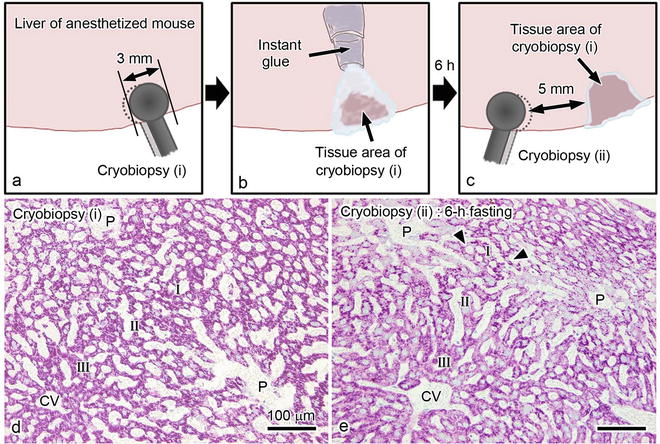
Fig. 51.2
Schematic representation of a series of cryobiopsy processes to obtain liver tissue specimens from anesthetized mice. A liver specimen is initially pinched off at one area with a pair of cryoforceps (cryobiopsy (i) in (a)), and bleeding is immediately stopped with an instant glue (b). After 6-h fasting, another liver tissue specimen is similarly obtained at a second area with the cryoforceps (cryobiopsy (ii) in (c)). (d) In the specimen of cryobiopsy (i), intense PAS staining is observed in almost all hepatocytes , the exception being those around portal vein s (P). (e) The PAS-staining intensity of the area of cryobiopsy (ii) is found to be decreased around portal tracts. Note the strongly PAS-positive hepatocytes remaining in parts of the middle of zone I (arrowheads). CV central vein , P portal vein. Bars: 100 μm
51.4 Detection of Different Consumption Under Local Circulation Loss Condition
To examine the effects of blood circulation on glycogen consumption, temporary stopping and reflowing of blood circulation were produced by binding with a circular clip and removing it (Fig. 51.3a). To prevent drying of the liver surface tissues, abdominal skin was quickly sutured, and a cotton sheet-absorbing physiological saline was placed on the mouse abdomen throughout the experiment. Temporary hypoxia of some parts of the clipped liver tissues had already been confirmed by measuring the oxygen saturation of similar liver tissues with confocal Raman cryomicroscopy , as previously reported [17]. After stopping blood circulation for 30 min and reflowing for 1 h, cryobiopsy of the restricted areas was performed on the inside (Fig. 51.3b-I) or outside (Fig. 51.3b-II) of the clipped liver tissues. In the liver tissues inside the clipped areas, the PAS -staining intensity was markedly decreased in certain areas around the portal vein s of zone I and also the central vein s (zone III) (Fig. 51.3c). Even in liver tissues outside the clipped areas (Fig. 51.3c, d), the PAS-staining intensity heterogeneously decreased as a mosaic pattern in all zonal areas. Thus, the PAS staining of liver tissues by cryobiopsy clearly revealed zonal difference s in glycogen storage after reflowing of blood circulation in living mice and probably reflects zonal metabolic functions in vivo.
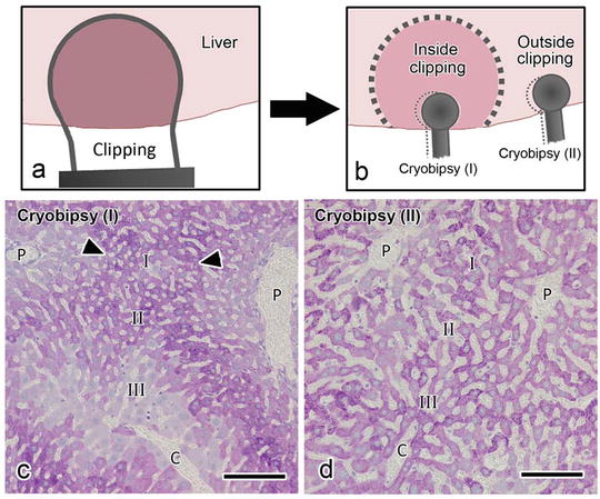
Fig. 51.3


Schematic representation of the experiment about anoxic blood circulation ; an area of the mouse liver tissue is clipped to stop the blood circulation for 30 min (a). One hour after removing the clip, some liver tissue specimens are obtained inside or outside the clipped area with cryoforceps (cryobiopsy (I) and (II) in (b)). Liver tissues were pinched off from inside ((c) cryobiopsy (I)) or outside ((d) cryobiopsy (II)) clipped areas, as shown in (b). (c) Marked reductions in PAS -staining intensity are detected in zone III and local areas of zone I near the portal tracts inside the clipped areas. Staining is still observed in areas far from the portal tracts in zone I (arrowheads). (d) The PAS-staining intensity is heterogeneously detected in all areas of the liver tissue outside the clipped area. CV central vein , P portal vein . Bars: 100 μm
Stay updated, free articles. Join our Telegram channel
Full access? Get Clinical Tree
