Hemodynamic Monitoring

Chapter 203 Hemodynamic Monitoring


BLOOD PRESSURE MONITORING


Blood pressure monitoring is extremely useful in critical cases. It allows fluid therapy to be tailored to a patient’s needs when used along with physical examination parameters, urine output, and even CVP monitoring. It is essential in guiding the use of inotropic agents and pressors, and these therapies should not be used unless blood pressure can and will be measured frequently. Normal blood pressure values for dogs are systolic pressures of 110 to 190 mm Hg and diastolic pressures of 55 to 110 mm Hg. For cats, normal ranges are 120 to 170 mm Hg for systolic and 70 to 120 mm Hg for diastolic. Mean arterial blood pressure (MAP) can be calculated from these measured values as follows:


image


Hypotension is defined as systolic blood pressure less than 80 mmHg or mean arterial pressure of less than 60 mmHg in either species. Causes of hypotension include decreased cardiac output secondary to reduced circulating volume, myocardial failure, severe bradyarrhythmia or tachyarrhythmia, or decreased systemic vascular resistance due to peripheral vasodilation secondary to sepsis or systemic inflammatory response syndrome. Treatment of hypotension should always be aimed at correcting the underlying problem (see Chapter 6, Hypotension).


Hypertension can be primary (essential hypertension), which is rare in both cats and dogs, or secondary from another disease process that alters renal or neurohormonal function. Renal failure, acute or chronic, is the most frequent cause of secondary hypertension, but hyperthyroidism, diabetes mellitus, hyperadrenocorticism, pheochromocytoma, and various medications (glucocorticoids, cyclosporin A, phenylpropanolamine, and erythropoietin) have also been associated with hypertension.


Blood pressure monitoring can be divided into two main categories, noninvasive and invasive methods. The noninvasive methods are used most commonly, and in veterinary patients they usually consist of either the oscillometric or Doppler methods, although photoplethysmography is also available. Invasive blood pressure monitoring provides direct arterial pressure measurement and is the most accurate method available.


Noninvasive Blood Pressure Monitoring


Noninvasive blood pressure monitoring is based on inflation of a cuff to occlude arterial flow, followed by measurement of the pressure at which flow returns. These methods are technically easy to use and require relatively inexpensive equipment but are prone to error, usually due to selection of an inappropriate cuff size. The guideline for the cuff width is approximately 40% of the circumference of the limb for dogs and 30% of the circumference of the limb for cats. If the cuff is too small, a falsely high pressure will be obtained; if the cuff is too large, a falsely low reading will result.1


The Doppler method measures only systolic pressure and usually is used in smaller animals such as cats, very small dogs, and exotic species. It is also useful in patients with hypotension or those that have arrhythmias because the Dinamap (device for indirect noninvasive automatic mean arterial pressure) is commonly inaccurate or does not give any readings at all in these circumstances. The Doppler method uses a 10-MHz ultrasound probe to detect blood flow in an artery. The probe is placed over an artery distal to the cuff. Doppler sounds become audible when pressure in the cuff is less than the pressure in the artery. Although the Doppler typically is regarded as measuring the systolic pressure, one study that compared Doppler readings with direct blood pressure monitoring in anesthetized cats found that the Doppler consistently underestimated systolic pressures by 10 to 15 mm Hg and was more closely correlated to MAP. This study was only in anesthetized healthy cats, so limitations are present.2


The Dinamap uses an oscillometric method of blood pressure determination. The cuff is alternately inflated and deflated, and during deflation alterations in cuff pressure are sensed by the transducer. These oscillations are caused by pulses in the limb. The peak amplitude of oscillations equals the mean arterial pressure. Systolic pressure equals the pressure at which oscillations are first detected, and diastolic pressure equals the pressure at which oscillations decrease rapidly.


Many machines calculate systolic and diastolic blood pressure from the mean arterial pressure using built-in algorithms, making the mean arterial pressure the most accurate value. The heart rate is measured as the number of oscillations occurring per minute, and should always be compared with the patient’s heart rate as determined manually or by ECG. The Dinamap has been evaluated in anesthetized and awake dogs and found to have reasonable accuracy in the normotensive ranges.3–6 The Dinamap is not reliable in cats7 or very small dogs, and even in appropriately sized patients errors can result if the patient moves or if significant arrhythmias are present. A similar device, the Cardell, also determines systolic, diastolic, and mean pressures and has been accurate in cats as well as dogs, eliminating some of the concerns associated with the Dinamap.8


Invasive Blood Pressure Monitoring


Invasive or direct arterial blood pressure monitoring is considered the gold standard for blood pressure measurement in both veterinary and human patients, awake and anesthetized. It is usually performed after inserting an arterial catheter that is connected to a pressure transducer and monitor, allowing for continuous monitoring of systolic, diastolic, and mean pressures. Techniques for direct arterial puncture and single-pressure measurement have also been described.


When a display monitor is employed, continuous direct arterial pressure monitoring allows for observation of pressure changes and trends (Figure 203-1). Another advantage of an arterial catheter is that it can be used to obtain blood samples for arterial blood gas analysis and laboratory testing. Despite its many advantages, direct monitoring should be limited to critically ill patients that will benefit from having their blood pressure measured frequently over a defined period, such as during anesthesia in a patient with a high anesthetic risk or while hospitalized in an intensive care unit.


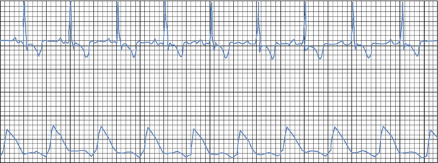
Direct arterial blood pressure monitoring in patients with hypovolemic or septic shock is extremely helpful in guiding volume replacement and the use of pressors to maintain an acceptable systemic blood pressure. By evaluating the pressure waveform with various arrhythmias, the clinician can distinguish which ones are causing poor pressures or even pulse deficits, and this can influence the decision as to whether or not to initiate treatment. Direct arterial blood pressure monitoring is not indicated in active, relatively healthy patients because of possible morbidity from arterial catheter placement and risk of the patient pulling the catheter out or disconnecting the arterial line and causing significant hemorrhage. Animals with arterial catheters must be strictly supervised at all times (see Chapter 49, Arterial Catheterization).


Once an arterial catheter is placed, it is connected to semirigid tubing that has been primed with heparinized saline from a bag of 0.9% sodium chloride with 1 unit of heparin per milliliter of saline. The fluid bag is pressurized to 300 mm Hg to prevent backward flow of arterial blood into the tubing. The tubing from the catheter is attached to a pressure transducer that is connected to a cable and mounted on a board placed at the level of the patient’s heart. The pressure transducer converts the pressure changes into an electrical signal that is carried to the monitor by the transducer cable, and then the signal is amplified and displayed on a monitor as a pressure waveform, showing the peak systolic pressure, dicrotic notch (which is created by closure of the aortic valve), and diastolic pressure. Monitors will also display numeric values for the systolic, diastolic, and mean arterial pressures.


Although direct arterial monitoring is considered the gold standard for blood pressure monitoring, it can give erroneous results if compliant tubing is used, the catheter is lodged up against the arterial wall, a clot forms at the tip of the catheter, air bubbles are present in the catheter or tubing, or the catheter or tubing becomes kinked. All of these problems can result in the waveform becoming damped, giving lower systolic and higher diastolic values than are present. Direct arterial blood pressure monitoring has higher associated morbidity than do noninvasive methods, including hematoma formation at the site of arterial puncture, infection, thrombosis of the artery, or necrosis of the tissues distal to the catheter (particularly in cats that have an indwelling catheter for more than 6 to 12 hours). Keeping the arterial line patent requires heparinization of the line and catheter, which can be of concern in very small patients. Fortunately, all of the complications other than hematoma formation are quite rare.

Stay updated, free articles. Join our Telegram channel

Sep 10, 2016 | Posted by in SMALL ANIMAL | Comments Off on Hemodynamic Monitoring

Full access? Get Clinical Tree

Get Clinical Tree app for offline access