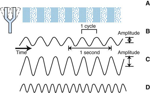
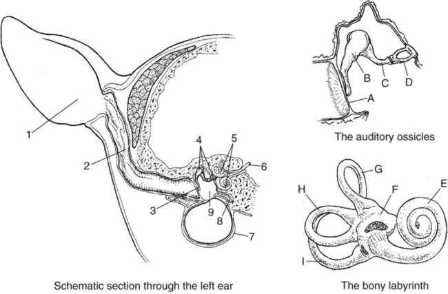
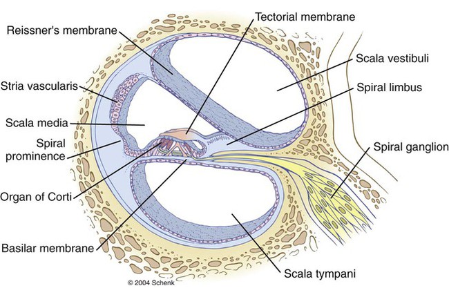
1. Sound waves are alternating phases of condensation and rarefaction (pressure waves) of molecules in the external environment. 2. Outer and middle ears funnel sound waves to the cochlea. 3. The cochlea is located in the inner ear. 4. The cochlea transduces sound waves to action potentials of the eighth cranial nerve. 5. Deciphering of sound wave frequency begins in the cochlea. 6. Action potentials from the cochlea are transmitted up through the brainstem to the cerebral cortex. 7. Deafness results from an interruption in the hearing process. Sound waves are longitudinal vibrations of molecules in the external environment characterized by alternating phases of condensation and rarefaction (increases and decreases in pressure). These alternating changes in pressure produce the sensation of sound after they strike the tympanic membrane and are subsequently transduced into neural signals that ultimately reach the cerebral cortex. Sound waves reaching the tympanic membrane can be expressed as changes in sound pressure as a function of time (Figure 17-1). The outer ear, composed of the fleshy part (pinna) and the ear canal, funnels sound waves to the tympanic membrane, or eardrum (Figure 17-2). Some animals can move the pinna to more effectively collect sound waves, and the natural shape of the pinna can act to selectively filter certain sound frequencies. The eardrum is a membrane between the outer and the middle ear. The middle ear is an air-filled cavity in the temporal bone and is connected to the nasopharynx by the auditory (eustachian) tube. Three tiny bones—the malleus, incus, and stapes—collectively called the ossicles, are connected to each other and are located in the middle ear. The malleus is connected to the eardrum, and the stapes is connected to the oval window, a membranous separation between the middle and the inner ear. The ossicles transfer vibration of the eardrum to the oval window in a manner that avoids a significant loss of energy as the sound wave is transferred from the air-filled outer ear to the fluid-filled inner ear. Two small skeletal muscles are also located in the middle ear, with one attached to the malleus and one attached to the stapes. Their contraction reduces the transfer of vibration between the eardrum and the oval window. This can function to protect the inner ear from very loud sounds. The inner ear (labyrinth) contains the receptor organs of two sensory systems: (1) the vestibular system, which detects acceleration and static tilt of the head (see Chapter 11), and (2) the auditory system, which detects and analyzes sound. The inner ear consists of the bony labyrinth and, within the bony labyrinth, the membranous labyrinth. The bony labyrinth is a series of tunnels within the petrous temporal bone. Inside these tunnels, surrounded by a fluid called perilymph, is the membranous labyrinth. The membranous labyrinth follows the contour of the bony labyrinth and contains endolymph. The vestibular and auditory portions of the inner ear are contiguous, and the “membranous tunnel within a bony tunnel” design is an anatomical feature of both parts. The auditory portion of this inner ear complex is called the cochlea (see Figure 11-1). The cochlear portion of the labyrinth is coiled like a snail shell. If we could mentally uncoil this arrangement to a linear form and then take a transverse section through it, perpendicular to the long axis (like cutting a salami and then looking at the cut end), we would see two membranes, the basilar and Reissner’s, dividing the cochlea into three chambers, or scalae (Figure 17-3). The dorsally located scala vestibuli and ventrally located scala tympani contain perilymph. The flexible middle scala, or scala media (cochlear duct), is formed by the membranous portion of the labyrinth and contains endolymph. The basilar membrane is the floor of the scala media, and atop this membrane lies the hair cell receptor organ for hearing called the organ of Corti. An anchored, gel-coated ridge, called the tectorial membrane, lies just atop the hair cells of the organ of Corti. The morphological organization just described is virtually the same all along the length of the cochlea, except that the scala vestibuli and scala tympani connect with each other at the distal end (farthest from the oval window).
Hearing
Sound Waves Are Alternating Phases of Condensation and Rarefaction (Pressure Waves) of Molecules in the External Environment
Outer and Middle Ears Funnel Sound Waves to the Cochlea
The Cochlea Is Located in the Inner Ear
< div class='tao-gold-member'>
![]()
Stay updated, free articles. Join our Telegram channel

Full access? Get Clinical Tree


Hearing
Only gold members can continue reading. Log In or Register to continue
