37 Head and neck pruritus
INTRODUCTION
Pruritus, crusting and ulcerative dermatitis of the head and neck is a common reaction pattern in cats. This is a particularly distressing presentation for both the owner and cat, and is often fairly refractory to treatment. There are four recognized feline cutaneous reaction patterns that are essentially manifestations of pruritus. Head and neck pruritus is one such pattern, and the other three are symmetrical alopecia, miliary dermatitis and lesions of the eosinophilic granuloma complex (see Chapter 36). These reaction patterns are not diagnoses and common underlying causes include hypersensitivity disorders, ectoparasitic diseases and microbial infections, and a systematic approach is required to identify and correct the underlying disease. The diagnostic approach involves careful and thorough history taking, clinical examination and diagnostic investigations. The success of long-term management of the case will depend on the final diagnosis. In this case a staphylococcal infection and an adverse food reaction were found to be responsible for the ongoing self-induced lesions on the neck.
CASE HISTORY
History taking should establish evidence of systemic involvement, seasonality, contagion, zoonosis, diet, management and response to previous treatment, in particular antimicrobial or glucocorticoid therapy (see Chapter 1). Some conditions that affect the head and neck may not exhibit early pruritus, but it can occur as the condition progresses and other complications occur. Thus, it is helpful to know whether the condition initially started as pruritus with subsequent development of lesions or whether there were already skin lesions present before pruritus became evident suggesting a primary eruption.
The relevant history in this case was as follows:
CLINICAL EXAMINATION
The relevant findings on examination in this case were:
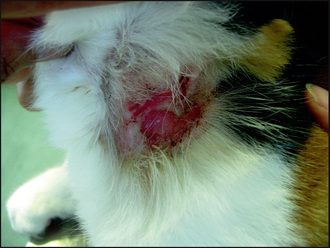
Figure 37.1 Self-induced excoriations, ulceration, erythema, crusting and alopecia on the lateral aspect of the neck.