43 Glaucoma – introduction
Glaucoma is the term given to the group of diseases in which a raised intraocular pressure leads to damage to the optic nerve and the retinal cells. It is a painful, often blinding, condition and is a very serious disease which can be difficult to treat. There are many causes of glaucoma and the choice of treatment often depends on the cause, the duration of the disease and the potential visual outcome.
The maintenance of normal intraocular pressure depends on a balance between production and drainage of aqueous humour. The ciliary body produces aqueous from the posterior non-pigmented epithelial cells by a process of both active secretion and passive ultrafiltration, and it flows through the pupil into the anterior chamber. It drains out of the eye through the iridocorneal angle (drainage or filtration angle). This pathway is shown in Figure 43.1. Once in the drainage angle, the aqueous passes through the pectinate ligaments and into the uveal trabecular meshwork where it is absorbed into the venous plexus vessels and out into the episcleral and scleral drainage veins (Figure 43.2). There is a second route of aqueous drainage – the uveoscleral or unconventional pathway. Here aqueous is drained via the ciliary body interstitial cells into the suprachoroidal space and choroidal and scleral vessels. The percentage of aqueous following this alternative pathway varies between species – it is more important in the horse than in dogs for example.
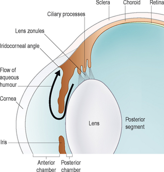
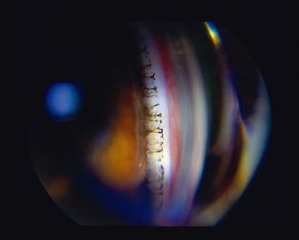
Figure 43.2 Drainage angle as seen on gonioscopy – the pectinate ligaments can be clearly seen spanning the angle.
Most cases of glaucoma result from impaired drainage rather than from overproduction. Tonometry, the measurement of intraocular pressure, is essential for the diagnosis and management of glaucoma. Normal intraocular pressures are listed in Table 43.1. Without the appropriate instruments to do this, it is impossible to diagnose and treat successfully, and therefore referral to an ophthalmologist is advised should a tonometer (Schiotz, Tonopen or TonoVet) not be available in the surgery. Since glaucoma is such a common ophthalmic problem, representing approximately 10% of ocular problems in one survey, it is strongly recommended that each veterinary practice has access to tonometry. In addition to measuring the intraocular pressure, gonioscopy – the examination of the iridocorneal (drainage) angle – is frequently necessary. The techniques for both tonometry and gonioscopy are discussed in Chapter 1. The latter technique requires practice to master and so is not routinely performed in general practice.
Table 43.1 Normal intraocular pressures in dogs, cats and rabbits
| Dog | Cat | Rabbit |
|---|---|---|
| 15–25 mmHg | 16–27 mmHg | 16–20 mmHg |
The clinical signs of glaucoma will vary depending on the species of animal, the cause, the speed of onset and the duration, together with how high the intraocular pressure has become. For ease of description the signs are usually divided into those seen with acute glaucoma and those in chronic cases – these are listed in Table 43.2. In general terms, the condition usually presents unilaterally and acute onset is common in dogs. Cats more frequently suffer from a chronic insidious onset. In dogs, pain, corneal oedema, episcleral congestion and blindness are the most consistent presenting signs whereas in cats it might just be a dilated pupil which is noted.
Table 43.2 Clinical signs of glaucoma
| Acute glaucoma | Chronic glaucoma |
|---|---|
Stay updated, free articles. Join our Telegram channel
Full access? Get Clinical Tree
 Get Clinical Tree app for offline access

|