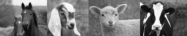
Angela M. Hughes, Kari J. Ekenstedt, Consulting Editors Inherited diseases and congenital defects are structural or functional abnormalities that may or may not be obvious at birth. There are a wide range of possible defects, from a single structural change (e.g., leg length) to the involvement of multiple organs or systems (e.g., storage diseases, chromosomal abnormalities), depending on the type and extent of toxic exposure during gestation or genetic mutation. It is important to identify animals with genetic or congenital disorders because of the economic and emotional impact on clients, but many disorders can be difficult to recognize and trace, particularly if they lead to embryonic or fetal death, abortion, dysmaturity, premature birth, or full-term stillbirth. It is important to remember that not all congenital defects are genetic in origin and making the distinction between conditions caused by an environmental factor and a genetic cause will help clients make appropriate changes to prevent the disorder in the future. To determine the cause of a disorder, it is vital that each case be examined thoroughly. Each case should be subjected to a full, careful description of the disorder, including a complete necropsy. The environment and management conditions should be investigated for potential causative agents, and all available genetic information for the affected animal(s) and unaffected herdmates should be collected, including sex, birth date, and breed. Environmental teratogens affecting large animals include toxic plants, drugs, viruses, and physical agents (e.g., hyperthermia, irradiation). Although it may be difficult to identify inciting teratogens, analysis of affected herds will reveal patterns that follow seasonal or management changes or stressful events. This chapter focuses on the essential genetic information that veterinarians require to inform their clients about genetic diseases and how to find information on diseases and traits with a genetic component. The basic blueprint for life in most organisms, the genetic material, is deoxyribonucleic acid (DNA). DNA is composed of two strands of nucleotides: adenine (A), thymine (T), guanine (G), and cytosine (C). These nucleotides, or bases, align to form complementary pairings such that A always pairs with T and G always pairs with C. These strands can be completely separated and replicated in preparation for cellular division. Alternatively, they can be partially separated temporarily, transcribed into ribonucleic acid (RNA), and translated into functional proteins. The DNA sequence of a mammalian genome is divided into autosomes and a pair of sex chromosomes, X and Y. Each chromosome contains a variety of genes as well as “filler” DNA that does not code for proteins, but may determine where and to what degree each gene is expressed. Genes on each chromosome code for the various structural and enzymatic proteins required for life. One set of chromosomes, and thus one set of genes, is inherited from each parent. As a result, a mammal generally has two copies, or alleles, of any given gene. Simple traits or diseases involving these genes can be inherited in dominant or recessive patterns and involve the autosomes or sex chromosomes. Unfortunately, not all traits or inherited diseases are simple; many involve multiple genes and interactions between genes and the environment, which can make it difficult to diagnose and understand a complex disease or trait. Traits or diseases with a recessive mode of inheritance require two affected alleles of the gene to express that trait or disease. These conditions can be autosomal, involving genes on the autosomes, or sex-linked, caused by genes on either the X or Y chromosome. Generally, autosomal recessive traits or diseases are caused by loss-of-function mutations in which the gene product has decreased ability or no ability to perform its original function. When an animal inherits the affected allele from both parents, it is homozygous for the gene and is unable to make functional copies of that specific protein. Heterozygous individuals inherit only one copy of the affected allele and they have a “backup” copy of the normal or wild-type allele to provide the functional protein. These heterozygous individuals are carriers of the trait or disease allele and can produce affected offspring if bred to another heterozygous or homozygous affected individual. The hallmarks of autosomal recessive traits include that all offspring of two affected parents are affected and that approximately equal numbers of males and females are affected. Unaffected carrier parents can produce affected offspring, and thus the trait or disease can skip generations. In addition, mating an affected individual to a noncarrier (homozygous normal) produces all offspring that appear normal but carry the affected allele. Breeding two carrier individuals results in approximately 25% normal, 50% carrier, and 25% affected offspring. An example of an autosomal recessive trait is spider lamb syndrome, or hereditary chondrodysplasia. Lambs affected with this syndrome display several skeletal abnormalities, including disproportionately long “spider” legs, curvature of the spine, facial deformities, rib and sternum deformities, lack of body fat, and muscular atrophy. The causative mutation is a single base-pair substitution, from a T to an A, altering a highly conserved amino acid in the fibroblast growth factor receptor 3 (FGFR-3) gene. This prevents the receptor from functioning to limit endochondral ossification and thus bone growth.1 Traits or diseases with a dominant mode of inheritance only require one affected allele to express the trait or disease, also known as the affected phenotype, and there is no distinguishable difference in the affected phenotype of a heterozygote and homozygote. Again, the genes that are responsible for these traits or diseases can be on the autosomes or sex chromosomes. Dominant traits or diseases may be caused by gain-of-function mutations that enable the mutant protein to have an altered structure or function. Alternatively, a single, functional copy of a gene may not be enough to achieve normal levels of function (“haploinsufficient”), resulting in the need for two normal copies of the gene to achieve a normal phenotype. The main feature of an autosomal dominant trait or disorder is that affected individuals must have at least one affected parent (unless it is a new mutation), and thus the disorder does not skip generations. There is no sex bias with an autosomal dominant trait, and approximately half the offspring of a heterozygous affected individual bred to a normal individual will be affected. Breeding two heterozygous affected individuals will result in 75% affected (one third of which are homozygous and two thirds of which are heterozygous) and 25% normal offspring. An example of an autosomal dominant disease is myotonia in goats. When startled or making sudden, forceful movements, these goats can develop severe, acute muscle stiffness causing immobility and sometimes falling over, resulting in descriptions such as “fainting,” “nervous,” “stiff-legged,” or “epileptic” goats. A single nucleotide change, from a G to a C, was identified that substituted a proline amino acid for a conserved alanine in a chloride channel in the muscle fibers. This alteration in the chloride channel causes a diminished channel-open probability at voltages near the resting membrane potential of skeletal muscle, resulting in decreased chloride conductance and a significantly decreased electrical threshold for firing action potentials. Ultimately, this altered chloride channel allows conduction of repetitive impulses that result in sustained muscle fiber contraction and stiffness.2 The co-dominant (or semi-dominant) pattern of inheritance is distinguished by the fact that homozygotes can be differentiated from heterozygotes based on clinical features. A well-known example of a co-dominant disease is hyperkalemic periodic paralysis (HYPP) in Quarter Horses. In HYPP the voltage-gated sodium channels in the muscle fibers have a mutation that increases sodium permeability across the skeletal muscle cell membrane. This causes increased muscle mass, but also drooling, prolapse of the nictitating membrane (“third eyelid”), respiratory stridor, and weakness. Homozygous HYPP horses experience more frequent and severe clinical signs of disease than heterozygous horses.3 Thus, it is important to counsel breeders to avoid producing homozygous affected horses by not mating two heterozygous horses, since approximately 25% of their offspring would be expected to be homozygous affected and 50% would be heterozygous, while only 25% would be homozygous normal. Breeding a heterozygous horse to an unaffected horse will also result in approximately 50% of the offspring being heterozygous with no possibility of producing a homozygous affected foal.
Genetic Disorders
Genetic Information
Recessive Inheritance
Dominant Inheritance
Co-dominant Inheritance
![]()
Stay updated, free articles. Join our Telegram channel

Full access? Get Clinical Tree


Genetic Disorders
Chapter 51
