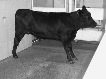
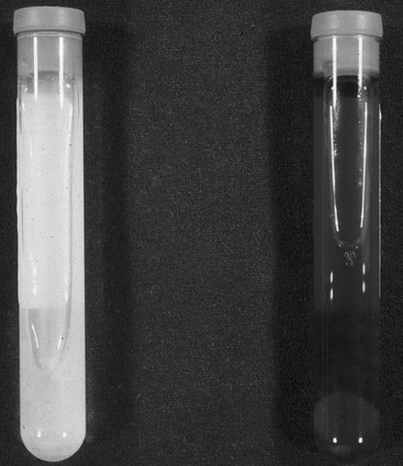
David C. Van Metre, Consulting Editor Dysuria is defined as difficult or painful urination. Stranguria is defined as straining to urinate, with the normal rate of voiding and urine egress being decreased and the effort required to void increased. Because these signs are often difficult to distinguish from each other and often present concurrently, they are considered together in this section. The most common causes of dysuria and stranguria are urethral obstruction, inflammation of the urethra and/or the bladder, and neurologic conditions that prevent normal emptying of the bladder. Adhesions between the bladder and other structures in the abdominal or pelvic cavities can create mechanical interference with bladder emptying, resulting in dysuria and stranguria. The horse voids urine actively and forcefully. Both male and female adult horses may briefly groan and strain slightly during normal urination. This should not be misinterpreted as dysuria or stranguria. In other large animal species, urination is more passive and straining or groaning normally is not observed. Conditions in which a horse may spuriously appear to have dysuria include lower urinary tract disease, abdominal pain, peritonitis, pleuritis, severe musculoskeletal disease, or neurologic disease. With any of these conditions, the horse may attempt to posture and urinate but may not be able to sufficiently increase intraabdominal pressure to allow complete voiding. Signs of dysuria and stranguria include treading, repetitive switching or flagging of the tail, pollakiuria (frequent voiding of small amounts of urine), flatulence during voiding, and retention of the urination posture for several seconds after voiding has ceased. Urine scalding of the perineal region or rear legs may be noted in either ruminants or horses with dysuria or stranguria. Vocalization during urination may accompany dysuria. While straining to void, the affected animal may show forceful contractions of the abdominal musculature. Male horses and ruminants that are experiencing dysuria or stranguria typically stand with the back slightly extended (mild lordosis), with the front legs held farther ahead of the body and the hind feet positioned farther behind the body than normal (Fig. 10-1). Dysuria or stranguria in large animals may be confused with tenesmus, or straining to defecate. This is most frequently a dilemma in neonatal foals with a ruptured bladder or meconium impaction. However, with tenesmus the rear feet of the foal are positioned slightly more anteriorly than with stranguria or dysuria. Stranguria may be severe enough in some cases to induce secondary rectal prolapse; therefore, when examining an animal with rectal prolapse, the clinician must establish whether or not the underlying cause might be urinary tract disease. Conversely, animals with tenesmus may strain to defecate with sufficient force to void small amounts of urine, and the observer may mistakenly attribute the problem to primary urinary tract disease. Urinary incontinence is defined as the involuntary voiding of urine. It is most frequently indicative of impaired neuromuscular control of urination. Concurrent fecal incontinence is commonly found if disease or trauma to the sacral segments of the spinal cord is present. Urinary incontinence may also occur with severe cases of lower urinary tract trauma and inflammation. In young animals, congenital abnormalities such as ectopic ureter must be considered in the differential diagnosis for urinary incontinence. Initially, the signalment, dietary and environmental history, onset of signs, duration, progression, and response to treatment should be established. Urethral calculi should be considered in castrated ruminants on high-grain diets. A history of one or more horses showing clinical signs of spinal cord disease, respiratory disease, stranguria, or urinary incontinence should immediately lead the practitioner to consider equine herpesvirus-1 myelitis in the differential. A history of dysuria or stranguria that develops after parturition usually indicates an injury to the lower urinary tract; such injuries can increase the female’s risk of subsequent urinary tract infection. For safety’s sake, the clinician should consider the potential for rabies as a primary cause before initiating the examination. A full physical examination should be performed because abnormal urination may be a sign of disease in other body systems, such as those characterized by diffuse muscular weakness. Common causes of dysuria, stranguria, and urinary incontinence are shown in Box 10-1. When possible, the animal should be observed urinating, and a sample of urine should be collected for dipstick urinalysis, measurement of specific gravity, and sediment examination; urine can be collected in a separate, sterile container for culture, if indicated. Urination can be induced in female cattle by gently rubbing the perineum immediately ventral to the vulva. In male cattle, the examiner may induce urination by placing a finger into the preputial cavity and gently rubbing the preputial mucosa. In ewes, urination can be induced by holding off the nose until the ewe struggles; urination typically occurs at this point. Obviously, this procedure should not be performed on ewes in shock or those with poor cardiac or respiratory function. In horses, goats, and male sheep, the examiner simply has to wait until the animal is ready to void, although urination may be encouraged by placing the animal in a freshly bedded stall. Recumbent animals will often void soon after standing. Normal equine urine is turbid owing to the presence of mucus and calcium-based crystals. The male’s preputial hairs and the female’s perineal region should be closely inspected for the presence of blood, exudate, or crystalline debris. Sedation and/or epidural anesthesia may be necessary to induce sufficient relaxation of the retractor penis muscles to enable examination of the penis. In prepubescent ruminants, the frenulum often prevents complete exteriorization of the penis for examination of the urethral orifice; general anesthesia may be necessary to induce sufficient relaxation. In bulls and steers, transrectal massage of the pelvic segment of the urethra may stimulate penile relaxation to enable penile visualization. The glans penis and urethral orifice should be carefully examined for masses such as papillomas, evidence of trauma, encircling hair rings, and embedded foreign bodies (e.g., grass awns). Penile examination is of particular importance in the dysuric or stranguric small ruminant because urinary calculi frequently become lodged in the urethral process (see Urolithiasis, Ruminants, Chapter 34). An accumulation of smegma, composed of mucus and cellular debris, may cause preputial swelling and dysuria in adult male horses. Smegma can usually be found as a hard, waxy mass in the urethral diverticulum. Preputial swelling without overt urinary dysfunction may be seen in equine Cushing syndrome (see Equine Pituitary Pars Intermedia Dysfunction, Chapter 41).1 In the male the penis and the urethra should be palpated percutaneously from the perineum distally to the sheath. Swelling, pain, abnormal urethral pulsations, and calculi lodged in the urethra may be detected. Urethral calculi are most commonly lodged just below the anus in male horses, and these can occasionally be palpated on the midline of the perineum. Marked swelling along the prepuce and ventral body wall in a bull or steer with active or recent dysuria/stranguria can indicate urethral rupture. The vulva, caudal vagina, and urethral orifice should be visualized and palpated in females. Sacrocaudal epidural anesthesia may facilitate examination if painful lesions are present. In females of breeding age, the cervix should be visualized or palpated and the uterus evaluated by palpation or ultrasonography because the pollakiuria and apparent dysuria that may occur at the onset of parturition may be the primary complaint of a novice observer. Previous dystocia can result in sufficient soft tissue trauma, laceration, swelling, and pelvic neuropraxia to induce dysuria or stranguria. The ventrum of the tail, perineum, udder, and hindlimbs should be examined for adherent blood or exudate originating from the female’s reproductive or urinary tract. In adult horses and cattle, rectal palpation should be performed when dysuria and stranguria are present. Before examination the clinician should take careful note of the tail and anal tone of the animal; reduction of either or both may indicate underlying neurologic or muscular disease. Introduction of the examiner’s hand and wrist into the rectum is usually sufficient for palpation of the pelvic segment of the urethra and bladder trigone. The caudal extent of the pelvic cavity should be carefully palpated for masses that might mechanically interfere with voiding. The bladder is typically located on the midline at the level of the pubic brim. Its presence in the caudal pelvic cavity, particularly in the standing animal, may suggest pelvic entrapment of the bladder. Bladder distention is commonly found in persistently recumbent horses and cattle, and on rectal examination the bladder is often positioned farther caudally than in standing animals. In the horse, bladder distention may also be found with abdominal or thoracic pain. Apparently, the abdominal pressure necessary to empty the bladder incites sufficient pain of diseased structures to cause reluctance to void. Musculoskeletal and neurologic disease may also result in bladder distention. These other possibilities should be carefully investigated when bladder distention is detected, yet no primary disease is found in the urinary tract. A careful rectal examination of the bladder and the proximal urethra of the horse might allow identification of urethral or cystic calculi. Most cystic calculi in the horse are singular and located in the trigone of the bladder and are palpable with the examiner’s arm inserted to the level of the wrist. If there is a large amount of urine in the bladder, the stone may not be palpable; in such cases, transrectal ultrasound examination may enable visualization of the stone. Sabulous calculi may be found in horses with stranguria or urinary incontinence, and on rectal examination the clinician may interpret the palpation findings as a bladder tumor or large stone.2 Detection of calculi in the bladder or urethra should prompt the clinician to consider the possibility of concurrent nephrolithiasis. If bladder dysfunction is not caused by structural abnormalities, trauma, or infectious disease, a thorough neurologic examination should be conducted. If neurologic dysfunction is suspected, an attempt should be made to determine whether the primary lesion is affecting the detrusor muscle or the urethral sphincter muscles of the bladder. This determination is often helpful in localizing the lesion and is important when selecting treatment. When bladder paralysis is caused by upper motor neuron (UMN) dysfunction, signs of UMN dysfunction may be evident in the rear limbs. The animal frequently postures and strains to urinate but voids only a small amount of urine because the striated urethral muscles are disinhibited from higher centers and their resultant increased tone impedes urine outflow from the bladder. Frequent, small-volume urine egress from the distended bladder occurs when the animal responds to the urge to void or when the bladder undergoes reflex contraction. With severe disease of the sacral spinal cord or sacral nerve plexus, lower motor neuron (LMN) input to the detrusor muscle is impaired or absent. Urinary incontinence is often the predominant clinical sign (e.g., cauda equina neuritis in horses or lymphoma in cattle). The bladder is usually moderately to severely distended, and urine can be expressed easily if pressure is applied to the bladder during rectal examination. With LMN dysfunction urine may also be voided as the animal walks. Voluntary or involuntary voiding is often incomplete, leading to retention of urine in the bladder. This, in turn, increases the patient’s risk of urinary tract infection and, in horses, sabulous calculi accumulation in the bladder. Other neurologic signs involving the sacral and coccygeal nerves may be apparent, such as decreased tail and anal tone and atrophy of the gluteal or tailhead musculature. Ataxia or weakness of the rear limbs may or may not be present with an LMN bladder. Urethral and bladder pressure profiles can be determined to better assess the location of the lesion.3–5 In small ruminants and neonates, transabdominal palpation is useful for evaluation of the urinary tract. In these animals a distended bladder can usually be palpated by simultaneously placing one hand on each side of the caudal ventral abdomen at the level of the pelvic brim and pressing the fingers of each hand toward the abdominal midline. If the bladder has been ruptured, it will be difficult to identify by palpation but ascites due to uroperitoneum can be detected. Digital rectal examination of the pelvic segment of the urethra can be performed in neonatal cattle and horses and in small ruminants. The umbilicus should be carefully palpated in neonates with dysuria or stranguria because urachal abscesses and adhesions to the bladder may impair voiding. An infected urachus will occasionally communicate with the bladder lumen, creating concurrent septic cystitis. Ectopic ureter(s) should be considered in young animals with persistent urinary incontinence; stranguria and dysuria are less common primary complaints. In affected females, vaginal urine pooling is often present. Vaginoscopic or cystoscopic examination can be performed, but the opening of the ectopic ureter can be difficult to locate during routine examination. Intravenous urography is typically required to locate the ectopic structure(s). As for all congenital defects, a careful assessment for defects in other organs should be performed in confirmed cases. If physical examination and urinalysis do not reveal the source of dysuria, stranguria, or incontinence, ultrasonographic evaluation of the urogenital tract should be performed. An endoscope can be used for visualization of the vaginal vault and preputial cavity and penis; air insufflation can be used to expand the walls and achieve a clear view of these structures. When advanced retrograde into the urethra, the urethral wall, bladder, and ureteral openings can be visualized. In neonates and small ruminants, plain abdominal radiographs, positive contrast urethrocystography, and intravenous urography are additional options. Hematuria is defined as blood in the urine. It may appear as occult blood detected during urinalysis, as uniformly red-colored urine throughout urination, or as blood clots passed at any phase of urination. If large clots are present, obstruction of the urinary tract may occur, resulting in concurrent stranguria and dysuria. Pigmenturia is defined as the presence of abnormal pigment in the urine; in large animals such pigments are usually limited to hemoglobin or myoglobin. Hemoglobin, myoglobin, and blood all cause a positive reaction for blood and protein on an orthotoluidine-based urine dipstick test. Certain oxidizing disinfectants can also trigger a positive reaction for blood on these strips. In addition, contamination of the urine with blood from the reproductive organs can result in hematuria, as can admixture of fecal blood with voided urine in females. Blood from penile or preputial injuries can contaminate the urine of males. When clots of blood are visible in the urine, the presence of hematuria is confirmed. Also, when scattered spots of color change are evident on the blood reagent pad of the urine dipstick, the reaction pattern reflects the presence of small aggregates of red cells deposited on the pad. Otherwise, differentiation of hematuria from hemoglobinuria and myoglobinuria requires that the discolored urine be centrifuged and the sediment examined. Hematuria is characterized by red-, pink-, or brown-colored urine that clears partially or entirely after centrifugation, resulting in a pigmented sediment pellet. With hematuria, red cells or red cell “ghosts” (red cells devoid of pigment) are visible on microscopic examination of urine sediment. Urine containing hemoglobin is clear to dark red in color, depending on the concentration of hemoglobin in the sample. If visibly discolored, the urine sample with hemoglobinuria does not clear when centrifuged. Animals with hemoglobinuria may be experiencing intravascular hemolysis, which leads to passage of hemoglobin from the plasma into the tubular fluid of the nephrons. The serum or plasma of these animals may be pink in color. Mucous membrane pallor or icterus may be evident, and tachycardia and tachypnea are present when red cell destruction is rapid and extensive. The packed cell volume may be decreased at the time of initial examination, or it may decrease progressively over 12 to 24 hours of monitoring. Release of hemoglobin from red blood cells in the vasculature may result in elevation of the plasma and serum total protein concentration. The clinician should note that hemoglobin is potentially nephrotoxic; renal function and hydration should be monitored in such cases. Sediment findings vary according to the underlying disease, and hyaline or cellular casts may be seen if pigment nephropathy is present. Urine containing myoglobin may be of normal color; if present in high concentrations in a sample, it imparts a dark red to brown color to the urine (Fig. 10-2). If visibly discolored, urine containing myoglobin does not clear when centrifuged. Sediment findings are variable but can include hyaline or cellular casts if pigment nephropathy is present. Myoglobin can be differentiated from hemoglobin in urine through ammonium sulfate precipitation, electrophoresis, or spectroscopy. Animals with myoglobinuria have muscle necrosis or injury, which leads to release of myoglobin from damaged muscle cells into the plasma and renal tubular fluid. Extensive muscle trauma (e.g., dog attacks, trailer accidents) or primary myopathies can induce myoglobinuria. Affected animals may show abnormal stance or gait or other evidence of muscle swelling, pain, or weakness. The serum activity of the enzymes creatine phosphokinase (CPK), aspartate aminotransferase (AST), and lactate dehydrogenase (LDH) is variably increased, depending on the duration and severity of muscle injury. As for hemoglobin, the persistence of myoglobin in the tubular fluid of the nephrons can induce tubular necrosis.
Alterations in Urinary Function

Dysuria, Stranguria, and Incontinence
Approach to Diagnosis of Dysuria, Stranguria, and Incontinence
Hematuria and Pigmenturia
Alterations in Urinary Function
Chapter 10