48 Generalized progressive retinal atrophy
CLINICAL EXAMINATION
General clinical examination is usually unremarkable, but the ophthalmic examination is not! Dogs might be nervous in the waiting room and can bump into objects on the way into the consulting room. Alternatively, they might walk with their head down, nose to the ground, and have a slightly high stepping gait. Cats will be reluctant to come out of their basket and might put their paws out to feel the edges of the table. Menace responses are likely to be reduced, and in most cases pupillary light reflexes are slow and incomplete. Thus the pupils are likely to be dilated in room light and even with a bright light source full and rapid constriction is unlikely to occur. Cats will retain a better pupillary light reflex than dogs. Both the menace response and pupillary light reflexes should be checked in bright (photopic) and dim (scotopic) light. Reactions are likely to be markedly reduced in the latter. In addition, the dazzle reflex is frequently reduced – the patient probably will still react to the bright light, but less quickly and with less intensity than a normal animal.
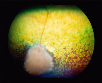
The retinal blood vessels will be narrowed and the optic disc can be small and pale (Figure 48.1). The non-tapetal fundus should not be forgotten, and the normal even pigment of the retinal pigment epithelium might be broken up into a patchy, ‘pavementing’ pattern. The changes in the retina are bilateral and symmetrical.
CASE WORK-UP
DNA tests are also available in some canine breeds to confirm the diagnosis and as a screening test for potential breeding stock. Blood or saliva samples can be used and results will indicate affected dogs, carriers and clear, unaffected animals. DNA tests are becoming available in more and more breeds and an up-to-date list of which breeds can be tested is available at www.optigen.com. In addition, both the Animal Health Trust and Cambridge University Veterinary School provide DNA tests for gPRA in some breeds.
Stay updated, free articles. Join our Telegram channel

Full access? Get Clinical Tree


