16 Gastrointestinal foreign body in a tortoise
Clinical Examination
As tortoise clinical examinations are restricted due to anatomical constraints, findings were brief:
DIFFERENTIAL DIAGNOSES
The differentials for reduced faecal output were:
The differentials for hindlimb paresis were:
Case Work-Up
Radiography
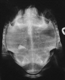
Three views were taken of the conscious tortoise – dorso-ventral (Fig. 16.1) using a vertical beam, and cranio-caudal and lateral using a horizontal beam. These orthogonal views outlined the presence of foreign material within the gastrointestinal tract. The object was less dense than bone but more radiodense than soft tissue, and its shape was consistent with a piece of gravel. The absence of gas near the mass suggested that it was not completely obstructing the gastrointestinal tract. No musculoskeletal pathology was detected on the radiographs.
Haematology and biochemistry
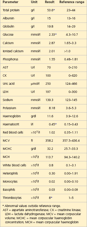
Financial consideration may preclude blood analysis in practice, and the clinician should be aware that analysis is not sensitive in reptiles. In this case, results showed mild dehydration (haematocrit and total protein were slightly elevated). There was no evidence of systemic infection (total white cell count normal, and no ‘toxic’ or ‘reactive’ white blood cells were seen on microscopy). Biochemical parameters (including renal parameters (uric acid), hepatic enzymes (AST, LDH, CK), and ionized calcium, and calcium to phosphorus ratio (1.85)) were within reported ranges – reducing the likelihood of metabolic bone disease or other metabolic derangement (Table 16.1).
Anatomy and Physiology Refresher
Gastrointestinal tract
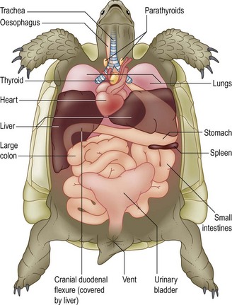
The chelonian oesophagus passes on the left-hand side of the neck (Fig. 16.2). The stomach is simple and fusiform, positioned caudal to the liver on the left with the pylorus centrally or to the right. Thick folds at the cardia in Testudo spp act like a sphincter; there may be a muscular sphincter at the pylorus. The small intestine is not well divided into sections, and lies caudally in the coelom. There is a muscular valve between the duodenum and caecum, caudally on the right. The large intestine consists of ascending, transverse and descending colon. The colon empties into the coprodeum of the cloaca, and thence material is voided via the vent. Various membranes and ligaments suspend the intestines, restricting movement of some parts of the gastrointestinal tract and permitting others to move more freely (e.g. the transverse colon can move dorso-ventrally, and heavy ingested material commonly becomes trapped in this region owing to gravity).
Stay updated, free articles. Join our Telegram channel

Full access? Get Clinical Tree


