Gastrointestinal Endoscopy: Instrumentation, Handling Technique, Training, and Implementation in Practice

Chapter 2 Gastrointestinal Endoscopy


Instrumentation, Handling Technique, Training, and Implementation in Practice


Gastrointestinal (GI) endoscopy is one of the best and yet most fundamental methods of examining the GI tract. It is a well-established procedure in veterinary medicine. The opportunity to examine directly and obtain tissue samples from the esophagus, stomach, and intestinal tract has greatly altered the clinical approach to diagnosis and made significantly more accurate the treatment of disorders of the digestive system. Despite the tremendous diagnostic advantages that endoscopy offers, it is still best used by the clinician as an adjunctive procedure in the evaluation of GI disease. A thorough history, physical examination, and selected laboratory and diagnostic imaging procedures (e.g., radiography and ultrasonography) as appropriate for each individual case are still important for thorough patient evaluation. When used judiciously, endoscopy offers a valuable alternative to exploratory surgery for direct examination of tissues, procurement of biopsy samples, retrieval of foreign bodies, and placement of gastric and jejunal (percutaneous gastrostomy-jejonostomy tube placement) feeding tubes.


Endoscopic equipment is no longer considered a luxury that only large referral centers or veterinarians practicing in affluent areas can justify purchasing. A variety of quality instruments, both new and used, are available. This chapter is concerned with key aspects to be considered in the purchase of an endoscope, the technical points of maneuvering an endoscope, steps toward gaining proficiency in operating an endoscope so that thorough examinations can be consistently performed (i.e., gaining proper training), and recommendations for increasing the use of endoscopy in the practice setting (recognizing indications for endoscopy and marketing the procedure successfully). Proper maintenance to promote the maximum life span of the equipment was discussed in Chapter 1. While there are now many small animal and mixed practices that have purchased endoscopes, there are also many practices that still vastly underutilize their equipment. Patient care can be clearly enhanced through earlier and more frequent use of endoscopy. Recognition of the many potential applications for the use of flexible endoscopy instrumentation is essential (Box 2-1), and success in fully implementing endoscopy services into the practice’s core procedures program depends on the veterinarian’s ability to effectively educate clients about the value of endoscopy as a diagnostic procedure and to make clear and effective recommendations on proper diagnostic assessment.


Subsequent chapters on the digestive system deal individually with the technical points of examining the esophagus, stomach, duodenum, ileum, and colon. The beginner learning to perform GI endoscopy naturally gives more thought to trying to move the instrument from one point to another than to careful gross examination and diagnosis of the areas being traversed. As the endoscopist becomes more skilled, an almost effortless maneuvering of the endoscope becomes second nature and the greater part of the time used to perform an examination is taken up with careful observation and synthesis of findings in relation to the clinical problem and prior experience. As the reader will find on review of the subsequent digestive system chapters, a combination of impressions from observation and microscopic review of biopsy samples is often needed for definitive diagnosis (see Chapter 8 for details on gross assessment and pathology). Because many disorders affect the upper and lower GI tract, a variety of appearances may be seen with an endoscope. The GI chapters carefully review the technical points of maneuvering the endoscope through these areas and provide an atlas of the things to be “seen” by the endoscopist. Indeed, upper GI endoscopy and colonoscopy are a primary means of physical diagnosis for the gastroenterologist.


The Decision to Purchase an Endoscope


For the veterinarian, the selection of equipment to be used for performing endoscopy often depends on its versatility of application, durability, and expense. Many practices, recognizing the full range of capabilities of endoscopy, have been able to financially justify the purchase of high-quality endoscopy equipment. A single scope can be used for a variety of GI and respiratory procedures. In addition, the availability of an endoscope often allows earlier access to examination of the GI tract than if surgery is the only other alternative. Clients almost always opt to have a less invasive procedure performed if the capabilities are present, and they often consent to this type of procedure much sooner than they would to surgery. Therefore, when consideration is given to the purchase of an endoscope, the most important factors to be reviewed should be the probable frequency of usage and versatility of the endoscope rather than the purchase price. Other important considerations are the quality of the optical system, the length and diameter of the insertion tube, the diameter of the instrument channel, and ease of operating the endoscope. Significant differences exist! Too frequently veterinarians rank a lower purchase price as one of the most important factors. This can be a significant mistake because even the most skilled endoscopist may find performing a complete examination and making the correct diagnosis difficult while using an endoscope of poor quality. High-quality endoscopy equipment will pay for itself in most practices in 1 to 3 years, as long as it is used as often as it can and should be. With proper care an endoscope should last many more years.


Training


Once the decision is made to purchase an endoscope, whether new or used, every effort should be made to become proficient in its use. Internal medicine residency training programs include extensive training in endoscopy and many opportunities to perform procedures under the guidance of experienced faculty through the course of the program (usually 3 years). The learning curve for general practice veterinarians in a private clinical practice setting is more challenging; nonetheless, learning the basic skills can best be accomplished through attending at least several formal wet lab courses and then practicing the skills of maneuvering an endoscope and procuring biopsy samples within a limited time period after the courses while the training information is still freshly in mind. Look for courses that offer at least several consecutive days of formal lecture and laboratory skills training. Single-day introductory classes are offered at some of the large national or regional veterinary conferences. Although these provide good introductory level experience, there simply is not enough time with the instructor(s) to gain the necessary endoscope handling skills in a single-day class. That is why it is best to enroll in a much more in-depth course where there will be plenty of laboratory time to repeat newly learned skills on a variety of dogs (repetition is extremely important) and also to learn upper GI endoscopy in cats. I also advise that, if more than one veterinarian at a practice will be performing endoscopic procedures, it is best that each attend formal training courses rather than a practice relying on one generalist who has attended a course to then train others.


Skills training should include proper handling techniques, maneuvering the endoscope through the upper GI tract smoothly and efficiently, techniques for traversing the pyloric canal to enter the duodenum, colonoscopy and ileoscopy, biopsy techniques, and foreign body retrieval. There is significant value in taking another course to fine-tune the necessary skill set after a period of time spent working on cases at one’s practice. This is an excellent opportunity to work with experienced instructors to correct any “bad habits” that may have been picked up and to simply continue practicing with the aid of experienced coaches. Endoscopy courses are advertised through university websites, brochures, seminars, and some of the endoscope company websites (e.g., www.lifelearn.com, www.ksvea.com, www.vetmed.wsu.edu, www.vet.uga.edu, www.cvmbs.colostate.edu).


Textbooks offer detailed descriptions of procedures and an array of normal and abnormal appearances for veterinarians to refer to in their own practice setting. Training CDs are also available (e.g., www.lifelearn.com). Please see the companion website for a demonstration of normal exam technique.


www.tamssmallanimalendoscopy.com


As is the case with successfully mastering any technical procedure, frequent practice definitely makes a significant difference in learning endoscopy. If proper skills of maneuvering, observation, and sample procurement are not developed, even the most sophisticated endoscopes are of little value and examination quality will be inferior. Frustration resulting from unfamiliarity with proper instrument handling and unavailability of necessary ancillary equipment too often lead to disuse.


Successful Implementation and Marketing of Endoscopy Services


For any new medical service to be successfully implemented, the medical staff needs to become properly trained in the techniques (builds confidence); and the support staff needs to be included in training and focus on the value they provide in assisting with the procedures and educating clients (engenders enthusiasm), thoroughly understand the value of the medical services offered, and ensure that clients are educated about the various diagnostic options available for their pets. If the doctors and support staff fully believe in the value of medical services and are enthusiastic about educating and encouraging clients, success is achievable!


The main reasons practices that have endoscopy equipment do not use the instruments more frequently are failure to recognize and appreciate the many indications for endoscopic examination, a lack of expertise in performing the procedures, being too conservative in recommending earlier use of more advanced diagnostic techniques, and not marketing the medical services to clients with more enthusiasm and conviction.


There are many indications for performing GI endoscopy in dogs and cats (Box 2-2), and these are discussed in subsequent chapters. A simple but often overlooked example of not recognizing indications is the need to check for esophagitis in patients with frequent vomiting. For example, some dogs and cats with a linear intestinal foreign body will develop esophagitis, sometimes of a moderate to severe degree, secondary to frequent vomiting of activated enzymes, acid, and toxins from the GI tract (the same can occur in animals vomiting frequently from any cause, including parvovirus enteritis and pancreatitis). Esophagitis causes significant pain, and significant injury can also result in fibrosis and even stricture formation (see Chapter 3).


Veterinarians are at a disadvantage compared with physicians when faced with determining whether a patient may have esophagitis. Physicians rely on clinical signs described by the patient. Veterinarians do not have this luxury. Many animal patients with esophagitis are missed because clinicians do not consider the possibility of gastroesophageal reflux disease (GERD) and because our patients are simply unable to “tell us.” Therefore, I recommend that endoscopy be done routinely at practices that have endoscopy equipment so that the esophagus and entire stomach can be examined immediately before any surgery for linear intestinal foreign body removal and any time an exploratory laparotomy is done on a patient with a history of unexplained vomiting. It is important to examine the stomach endoscopically in this situation because it is not possible to examine it entirely during exploratory laparotomy. This endoscopic examination of the esophagus and stomach can be done easily at any hospital that has GI endoscopy equipment; once proper experience is gained it only takes a minute or two to thoroughly examine the esophagus and a few more minutes to evaluate the stomach. In this way a patient that has esophagitis is recognized early, and appropriate therapy can be instituted along with any surveillance that will be necessary during the ensuing weeks for any animal that has significant esophageal injury. In addition, patient care is clearly enhanced. A quick endoscopic examination of the esophagus and stomach done before surgery is charged at a reasonable fee and somewhat lower than what a full examination (including biopsies) would cost.


One of the most important advantages of endoscopy is that it allows early examination of the GI tract. Too often patients with GI disorders do not have a definitive diagnosis established soon enough. It is still important to perform basic diagnostic tests and consider therapeutic dietary trials early in the clinical course, but sometimes the preliminary assessment coupled with various trial therapies goes on for far too long before more definitive diagnostic steps are taken. Although pet owners may be initially reluctant to consider a procedure like endoscopy, usually because of concerns related to the requirement for anesthesia or cost constraints, an important part of the marketing of endoscopy is to educate clients about the value of early examination, especially in patients with intermittent or early chronic vomiting (10 to 14 days or longer). Of course, routine baseline tests should be done first as part of the initial screening diagnostic plan (e.g., a complete blood count, complete biochemical profile, urinalysis, fecal examination [centrifugal flotation combined with a Giardia antigen test], abdominal radiographs, and dietary trials, as deemed appropriate). When a definitive diagnosis is established early it not only can decrease overall costs to the client but also, very importantly, can effect better patient care through an earlier determination of specific therapy for the patient’s condition. Further, endoscopy is a very successful method for removing foreign bodies from the upper GI tract of dogs and cats; its success rate is greater than 80% (see Chapter 7 for details on foreign body retrieval). Clients naturally are very pleased when foreign bodies can be removed via endoscopy at less cost and in a much less invasive way as compared with surgery.


Besides the value of early diagnosis, another key feature of endoscopy is that it is safe and minimally invasive. The anesthetic risk factors of each individual patient are the only consideration. Clients almost always opt to have a less invasive procedure performed if the capabilities are present, and they often consent to this type of procedure much sooner than they would to surgery.


Finally, other marketing techniques can include providing clients with a few representative photos from their pet’s procedure (see Chapter 1 for a discussion on image capture devices), using a library of photos (e.g., textbook atlas photos or photos from the hospital’s files) to explain the value of endoscopy, providing client handouts describing endoscopy for animals, and illustrating case reports in client newsletters or on examination room walls. The bottom line is that when the doctors and staff make recommendations with enthusiasm and conviction, the number of procedures that are done will increase significantly. For hospitals that do not offer endoscopy, this section provides many reasons to consider referral for endoscopy.


Selection of Endoscopic Instruments: Making the Correct Choice


Many types of endoscopes are available. A standard upper GI endoscope suitable for esophagogastroduodenoscopy (EGD) in dogs and cats should be a minimum of 100 cm long (working length) and have four-way distal tip deflection with at least 180-degree upward deflection, water flushing, air insufflation and suction capabilities, independent locking deflection controls, an accessory (instrument) channel with a diameter of 2 mm or greater, and forward-viewing optics. Most newer endoscopes now feature an upper deflection capability of 210 degrees, and the latest model endoscopes made specifically for use in small animals have insertion tube lengths of 140 cm. Newer instruments that are fluid tight and immersible are easier to clean.


Veterinarians are cautioned against purchasing endoscopes with two-way, rather than four-way, distal tip deflection capability for use in examining the GI tract. Many of these endoscopes are also relatively short (50 to 80 cm) and were originally manufactured as bronchoscopes. Although often less expensive (especially when sold as used equipment), these endoscopes are not versatile enough to facilitate a smooth and thorough examination of the stomach, antral canal, duodenum, and colon. A complete examination can be performed in some cases, but maneuvering through the antral canal, pylorus, and duodenum is considerably more difficult with a two-way rather than a four-way endoscope. Also, the shorter length precludes duodenal examination in many dogs, which is important in all cases in which vomiting, diarrhea, or weight loss is part of the clinical presentation. The ability to routinely perform a thorough examination of the stomach and descending duodenum in the majority of patients who undergo endoscopy is well worth the added expense of a more versatile endoscope.


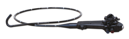
Careful thought must be given to the insertion tube diameter and length of the endoscope. Until recently, most endoscopes used in veterinary practice were designed for use in humans and often classified as adult or pediatric. Insertion tube diameters of endoscopes manufactured for examination of the human upper GI tract range from 6.0 (pediatric size) to 12.8 mm and often have a working length of around 100 cm. Fiberscopes manufactured for veterinary use are available in an insertion tube diameter size as small as 7.8 mm. Smaller diameter video endoscopes with larger instrument channels (up to 2.8 mm) are now available (see Chapter 1 for mechanical details on fiberscopes and video endoscopes). Most endoscopes used currently in small animal practice are in the range of 7.8 to 9.8 mm (Figure 2-1), and even smaller diameter video endoscopes will be available in the future. The inner working channel diameter of the human and veterinary endoscopes varies based on insertion tube diameter but ranges from 2 to 2.8 mm.


Veterinarians purchasing their first endoscope should consider a single high-quality endoscope that may be used for both upper (EGD) and lower (colonoscopy and ileoscopy) GI endoscopy in cats and small dogs, in which scope diameter is a key consideration, and in larger breed dogs, in which scope length is a more important consideration. Although specialty hospitals will generally have a variety of endoscopes available to facilitate all types of examinations in the smallest to largest of small animal patients, it is not feasible for most general practices to purchase more than one to two flexible endoscopes. Thus, for general practices, the key consideration is focused on the versatility of a single scope. Referral to a well-equipped specialty center should be considered in situations in which the available scope is not suitable for the size of the patient. GI scopes can also sometimes be used for bronchoscopy procedures in medium to large size dogs. Human pediatric scopes have an excellent diameter for small animal patients, but their length of 100 cm is too short to reach the duodenum of many larger dogs. This, however, is an excellent scope for feline-only practices (100-cm working length and 7.8-mm diameter; e.g., Olympus XP-20).


The insertion tube diameter should range from 7.8 to 9.8 mm. The major limitation of a larger insertion tube (greater than 9 mm) is that passing it through the pyloric canal to the duodenum in cats and small dogs is more difficult. This is a function of both the level of expertise of the endoscopist and the size of the pyloric canal. Even experts will be unable to advance a 9.8-mm endoscope through the pylorus of some cats. Great care must be taken when attempting to pass a scope through a narrow pylorus, expecially in cats, because it is possible to cause a perforation in the stomach if too much force is exerted in a tight canal. The endoscopist who acquires adequate maneuvering skills will be able to routinely traverse the pyloric canal to enter the duodenum, even in most animals as small as 1.4 to 1.8 kg (3 to 4 lbs), when using a smaller diameter endoscope (7.8 to 9.0 mm). The larger endoscopes can be used effectively in many animals, but a complete examination in very small patients is inherently more difficult. This becomes an important consideration for any urban practice in which many feline and small canine patients are seen.


What is sacrificed when a pediatric or small animal veterinary endoscope is purchased rather than a larger diameter endoscope? Pediatric endoscopes have instrument channels of 2 to 2.5 mm, whereas the larger units often have instrument channels with diameters up to 2.8 mm. This is changing, however, with newer technology video endoscopes that have a larger instrument channel in a small-diameter insertion tube. The larger instrument channel (2.5 mm to 2.8 mm) allows for the use of suction while biopsy forceps or another instrument is present in the channel. Simultaneous suction with a smaller working channel is not as effective; the instrument usually needs to be withdrawn from the channel before suction can be used to remove a large amount of air or fluid. However, this is not a significant drawback because simultaneous use of instruments and suction is not usually required, except during some procedures for retrieval of gastric foreign bodies. The smaller working channel does not accommodate certain accessory instruments such as larger biopsy instruments and some of the larger foreign body graspers. However, other instruments can be used effectively for biopsy and foreign body removal, so these limitations are quite minor. No substitute exists for a thorough examination, and the pediatric endoscope offers this capability to the endoscopist better than any other type of endoscope. With the use of proper instruments for biopsy and foreign body retrieval in addition to good ancillary instrument technique, endoscopes with 2-mm biopsy channels still work well.


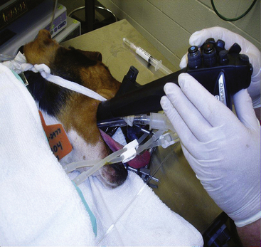
The ideal working length of an endoscope for small animal practice is 140 cm because it provides sufficient length to reach the duodenum in the largest of dogs. A 100-cm scope, the type that is still commonly found in general small animal practices, is too short to reach the duodenum of larger dogs, which could include any dog weighing more than 27 kg (60 lbs). A 100-cm scope can actually be long enough to reach the duodenum of some dogs weighing 35 to 40 kg (77 to 90 lbs), but in dogs of certain breeds that weigh less, the length simply may be insufficient (Figure 2-2). There are significant differences among breeds, so for maximal versatility the longer scope is best. The 140-cm veterinary scopes have a small enough diameter (7.8 to 9 mm) that they can be used for duodenoscopy in most cats. Most specialty practices will maintain both a long scope and a shorter one of smaller diameter (e.g., 100 cm in length and 7.8 mm or less in diameter) to facilitate examination of the smallest cats and dogs. For practices that do not have a scope longer than 100 cm, referral should be considered if an upper small bowel examination will be needed for a large dog (Figure 2-3).


Stay updated, free articles. Join our Telegram channel

Sep 10, 2016 | Posted by in SMALL ANIMAL | Comments Off on Gastrointestinal Endoscopy: Instrumentation, Handling Technique, Training, and Implementation in Practice

Full access? Get Clinical Tree

Get Clinical Tree app for offline access