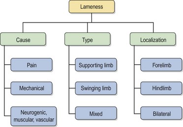
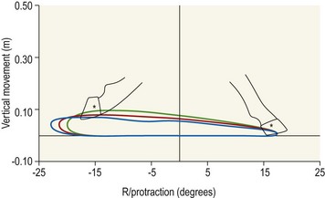
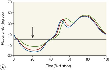
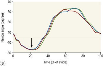
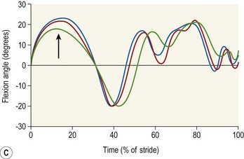
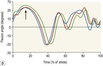
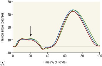
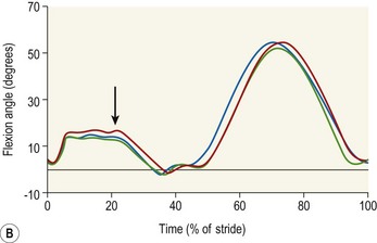
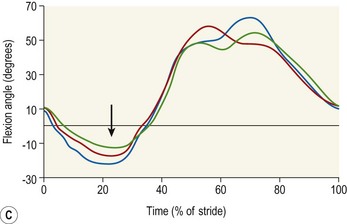
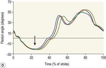
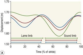
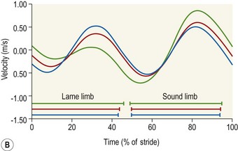
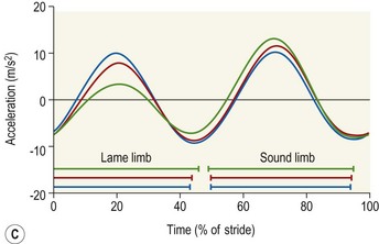
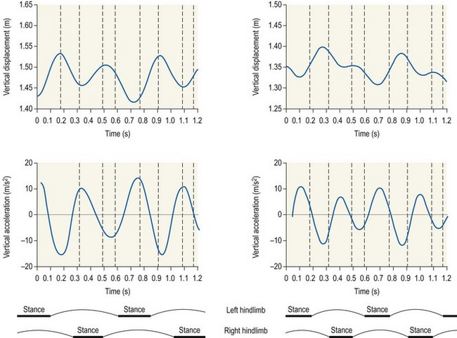
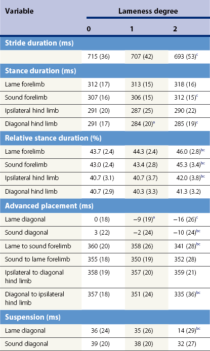
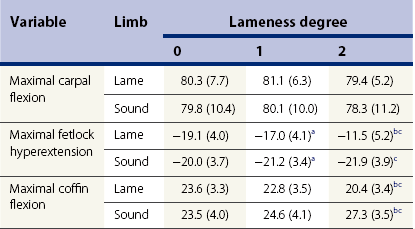
Chapter 9 Horses are kept as domestic animals due to their outstanding locomotor skills, which have been employed for military use, transportation and sports. Perfect athletic performance, of course, needs a sound locomotor system. Maintenance of soundness and detection of lameness are of prime importance for horse owners. The ability to study the gait of horses and assess the small deviations associated with locomotor problems is limited by the physiological ability of the human eye. Therefore, locomotion research in horses started very early with studies of the gait of lame horses. Already in 1899, Muybridge published series of photographic plates of both sound and lame horses in his fascinating book, Animals in Motion. These nice studies were the start of modern kinematic research about lameness in horses, which is and, will continue to be, a central theme of equine locomotion analysis (Leach & Crawford, 1983). Lameness is not a disease but a symptom of a locomotor disturbance. Generally, lameness can be defined as an alteration of the normal gait due to a functional or structural disorder in the locomotor system (Wittman, 1931; Knezevic, 1982; Stashak, 1987; Wyn-Jones, 1988; Speirs, 1994; Wilson & Keegan, 1995). A detailed description of the locomotion pattern is a central part of each lameness examination. The goal is to localize the cause of the lameness and to make a diagnosis that is as specific as possible as a basis for the veterinary therapy. In a clinical setting various classifications of lameness are used to differentiate various pathological gait patterns. Such classifications provide only a rough framework and a real lameness cannot always be fitted into a single category. Nevertheless, placing a lameness in one of the categories is a first step for the clinician in reaching a diagnosis. Locomotion analysis, on the other hand, should provide specific details of the various lamenesses as well as fundamental relations and principles. These can be used to understand the different types of lameness and to provide the scientific basis for interpreting the observations made during lameness examinations. Figure 9.1 shows the traditional classifications for lameness. The cause of the lameness is most often pain in one or more limbs. Sometimes diseases of peripheral nerves, blood vessels or muscles cause specific lameness and occasionally purely mechanical restrictions can be found. The type of lameness describes the phase of the stride when the disturbance is caused. A supporting limb lameness is caused by pain during the stance phase, while swinging limb lameness is caused by problems during the swing phase. Pure swinging limb lameness without pain during the stance phase is extremely rare and is most often a component of mixed lameness with features of both supporting and swinging limb lameness. The third classification describes the site of the lameness as a fore or hind limb lameness, or as a bilateral lameness when both fore limbs or both hind limbs are affected. If a specific structure can be proven as the origin of lameness and a specific diagnosis is established, locomotion analysis techniques can be used to define the associated locomotor patterns. A special situation is represented by induced lameness, which has been used in several clinical studies for evaluations of diagnostic or therapeutic regimes. Lameness models have been reported to induce hoof lameness (Merkens & Schamhardt, 1988a; Foreman & Lawrence, 1991; Keegan et al., 2000), arthritis of the carpal joint (Auer et al., 1980; Firth et al., 1987; Peloso et al., 1993) and tendonitis (Silver et al., 1983; Williams et al., 1984). The ability to induce a more or less transient lameness in groups of sound horses offers an important and reliable method to study the locomotor pattern in horses with specific, well-defined lameness in a controlled manner that minimizes individual variations. 1. Firstly, the kinematics that are specific to different types of lameness will be described. This will provide a more complete insight into the gait adaptations and how horses deal with and compensate for pain in a limb. The complete picture of movement changes of all body parts due to fore or hind limb lameness makes it possible to distinguish the original and compensatory lameness. The findings are of fundamental importance for the orthopedic specialist. 2. The effectiveness of the locomotor adaptations in terms of load reduction in the painful limb and load redistribution will be evaluated using kinetic methods. The connection of kinetic and kinematic findings allows an analysis of the mechanisms by which a horse manages a lameness. 3. The patterns of some well-defined lameness will be presented and analyzed to determine their specific signs, which will serve as a diagnostic database. 4. The possible use of locomotion analysis techniques in clinical settings, such as the documentation of diagnostic or therapeutic regimes or the diagnosis of insidious lameness, will then be described and discussed including the pros and cons of these methods. 5. Finally, the effects of several frequently used diagnostic or therapeutic aids will be evaluated. In these studies locomotion analysis technology is used to evaluate the proposed effects and to support or to reject their use based on scientific fundamentals. 1. A comparison with the ‘normal’ locomotion pattern in the population, which is based on the experience of the clinician. This requires a sufficient number of observations in sound horses to establish a standard and to eliminate the problem of individuality. This ‘standard horse’, however, may not be the best method for kinematic assessments due to the small differences caused by lameness compared to the wide interindividual variation in sound horses. 2. A comparison using the horse’s own pattern as an individual control. Since the sound pattern of the individual is usually unknown during lameness examinations, diagnostic nerve blocks serve as a tool to restore kinematics that are close to the sound pattern during a clinical evaluation. In a research setting similar comparisons are possible using lameness models, where the sound locomotion pattern of each horse before lameness induction serves as an individual control and minute deviations due to lameness can be easily differentiated from the small intraindividual variations. 3. An assessment of the movements of the left and right sides of the body in terms of locomotor symmetry or asymmetry within an individual horse. This approach allows an immediate comparison of affected and unaffected limbs independent of individual characteristics. Using one of these three methods several general characteristics of supporting limb lameness and swinging limb lameness have been described. Many studies (Hugelshofer, 1982; Clayton, 1986b; Girtler, 1988a,b,c; Tietje, 1992; Buchner et al., 1995a; Keegan et al., 1997; Weishaupt et al., 2006) have tried to find relationships between types of lameness and the timing of the limb placements, described as stride variables. However, these studies did not find typical changes or asymmetry patterns. This is in contradiction to subjective impressions, where some observers describe that a shortening of the loading phase of the lame limb seems to be a typical sign for a supporting limb lameness (Wittmann, 1931; Ratzlaff et al., 1982; Hajer et al., 1988). Nevertheless, some general temporal patterns can be seen if the lameness is moderate or severe. Slight or ‘subclinical’ lameness, on the other hand, does not show significant temporal deviations from the sound stride pattern (Buchner et al., 1995a; Weishaupt et al., 2006). Generally, lame horses tend to move with a slower velocity. This is of utmost importance for the interpretation of the stride pattern in lame horses, because all stride variables are heavily dependent on velocity (Dusek et al., 1970; Leach & Cymbaluk, 1986; Leach & Drevemo, 1991). Furthermore, the lameness degree is influenced by speed, as moderate lameness increases with speed (Peham et al., 2000). Comparisons of different assessments, for example before and after diagnostic anaesthesias, are valid only if recorded at the same velocity or using statistical correction methods (Kübber et al., 1994). Horses reduce their velocity by decreasing stride duration, stride length and all the dependent variables, such as stance duration or contralateral advanced placement. On the other hand, when horses are forced to maintain a constant velocity on the treadmill, variable results regarding stride duration and stride length in lame horses have been reported (Buchner et al., 1995a; Pollhammer-Zeilinger, 1996; Keegan et al., 1997). When showing different degrees of hoof lameness, provoked by the screw model of Merkens & Schamhardt (1988a), horses maintained the velocity by taking shorter, but quicker, strides than in the sound condition (Table 9.1) (Buchner et al., 1995a; Galisteo et al., 1997; Weishaupt et al., 2006). However, in horses suffering from navicular disease no consistent changes in stride duration and stride length were found between the lame pattern and the sound pattern after diagnostic anesthesia (Pollhammer-Zeilinger, 1996; Keegan et al., 1997). Table 9.1 Significant differences between the values of a variable for different lameness degrees are indicated by superscripts: a, degree 0 versus 1; b, degree 1 versus 2; c, degree 0 versus 2. Diagonal advanced placement is positive when the hind limb precedes the forelimb and vice versa. Reprinted with permission from: Buchner et al. (1995). Generally, a consistent feature in all studies is a shortening of the swing duration in lame horses compared to the same horses without lameness (Hugelshofer, 1982; Tietje, 1992; Buchner et al., 1995a; Keegan et al., 1997). In other words, horses lengthen the stance phase duration rather than shortening it to diminish the pain. This is in contradiction to the opinions that shortened stance durations are indicative of supporting limb lameness (Ratzlaff et al., 1982; Clayton, 1986a). Furthermore, there is no difference in stance duration between the lame and the sound limb (Girtler, 1988c; Tietje, 1992; Buchner et al., 1995a; Weishaupt et al., 2006). The horses maintain their symmetry; both limbs are kept on the ground longer with increasing lameness, while both swing durations decrease. This does not contradict the findings of differences in stance durations between left and right limbs in some particular horses. Absolute symmetry is nearly impossible in nature and significant left/right differences as sign of handedness or leggedness are found in various breeds (Drevemo et al., 1987; Deuel & Lawrence, 1987; Meij & Meij, 1980). Asymmetry in stance duration is therefore not a sign of a supporting limb lameness, but a typical individual characteristic. In forelimb lameness, a real asymmetry due to lameness can be found in the suspension phase at the trot (Table 9.1) (Clayton, 1986a; Buchner et al., 1995a; Weishaupt et al., 2006). The suspension phase following the lame diagonal stance phase, which means the time when none of the limbs is on the ground after the stance phase of the lame forelimb and diagonal hind limb, is significantly shortened in lame horses. This is a sign of reduced propulsion during the stance phase of the lame limb. The suspension following the sound diagonal, on the other hand, is nearly unchanged. This asymmetry can only be seen in forelimb lameness, not in hind limb lameness. The asymmetric suspension phase in forelimb lameness has some implications for the co-ordination of the placements of the different limbs. The duration of the step from the lame to the sound forelimb is shorter than the contralateral step or than the step duration without lameness (Table 9.1) as a result of the clearly shortened suspension between the lame and sound diagonals. An interesting pattern is seen in the diagonal advanced placement. As already mentioned, during forelimb lameness, the stance phases of the forelimbs tend to increase, with the fore hooves being placed earlier and lifted later. This results in an earlier placement of the forelimbs in relation to the diagonal hind limbs. Sound horses place their diagonal limb pairs almost synchronously or even place the hind limbs earlier than the forelimbs (positive diagonal advanced placement). The latter sequence may be indicative of superior gait quality in dressage horses (Holmström et al., 1994). Lameness reverses this pattern. Similarly, horses on a treadmill show this earlier forelimb placement, perhaps due to a need for longer ground contact, possibly as a sign of a remaining insecurity even after a long period of habituation to the treadmill (Buchner et al., 1994). During hind limb lameness, generally the same temporal pattern is found in most stride variables, however, the amount of changes in limb timing is much less in hind limb lameness. Stride frequency increases as in forelimb lameness, but the increase in relative stance duration is small and both diagonal advanced placement and suspension phases do not change at all (Buchner et al., 1995a). Horses with hind limb lameness keep this variable constant and perfectly symmetrical. There are two possible explanations for this temporal stability. Firstly, load damping may be more effective in the hind limbs as a result of greater tarsal flexion during loading of the lame limb. And secondly, a lower percentage of the body weight is carried by the hind limbs (46.8%, trot) compared with the forelimbs (53.2%) (Merkens et al., 1993), which may facilitate the management of a supporting limb lameness. Hoof movement can be visualized as the hoof trajectory during the stride. Figure 9.2 shows the trajectory of a fore hoof on a treadmill seen from the right side. The stance phase is characterized by a constant vertical position on the treadmill belt during the horizontal movement from right to left in the figure. After lift-off, the flight arc of the hoof shows the elevation during the swing phase and ends with the hoof landing at the start of the next stance phase. Different features can be seen in this figure showing typical changes in the hoof trajectory due to various degrees of forelimb lameness. The maximal height of the hoof during protraction of the limb is said to be lower during both supporting limb lameness and swinging limb lameness (Stashak, 1987). In supporting limb lameness, a lower flight arc might reduce pain on impact. In swinging limb lameness, difficulties in flexing the joints may cause a lowering of the flight arc. However, this could not be confirmed in recordings of horses suffering from navicular disease. Several authors investigated this lameness for characteristic patterns, but did not find consistent changes in maximal hoof height (Ratzlaff & Grant, 1986; Pollhammer-Zeilinger, 1996; Keegan et al., 1997). In experimentally induced forehoof lameness (Buchner et al., 1996a) as well as in patients with forelimb lameness (Girtler et al., 1987) a higher flight arc was found in the sound forelimb and an unchanged height in the lame limb (Fig. 9.2). During hind limb lameness, a lower flight arc was found in the lame limb, while the sound limb had an unchanged flight arc (Buchner et al., 1996a). Both patterns give the same impression, the hoof of the lame side is lower than the contralateral hoof. In induced toe pain lameness, a shift of the instant of maximal hoof height nearer to midswing has been found (Keegan et al., 2000). Changes in the pro- and retraction of the limbs of lame horses are visible at the walk. Retraction of the forelimbs is slightly reduced during forelimb lameness. For the interpretation of this pattern a comparison with the changes in hind limb movement during hind limb lameness is interesting. Lame hind limbs show a reduction in protraction rather than retraction. Perhaps the position of the limb relative to the body center of mass influences this feature. During walking vertical ground reaction forces of the forelimbs reach peak values in the second half of the stance phase, when the limb is retracted, which brings it closer to the body center of mass. On the other hand, the position of the hind limbs relative to the body center of mass causes peak loading in the first half of the stance phase (Merkens et al., 1986). The changes in pro- or retraction, which are more obvious during walking than trotting, might reduce the total load on the lame limb by shortening the period of high load. The changes in the temporal stride pattern also have implications for the linear stride variables. The distances between hoof placements of sound and lame limbs and vice versa, which are called the step lengths, might give information about the cause of the lameness. A shortening of the step length from the lame to the sound limb is said to be typical for a supporting limb lameness (Wittmann, 1931; Stashak, 1987). Horses with sesamoiditis (Clayton, 1986b) and hoof lameness (Buchner et al., 1996a) showed this feature. This shortening corresponds with the temporal variables in terms of the shorter advanced placement between lame and sound forelimbs as well as the clearly shortened suspension phase following the lame diagonal. The linear stride variables offer further proof of reduced propulsion during the stance phase of the painful limb. Few studies have described specific changes in the hoof-landing pattern due to lameness. Toe first or heel first might give information about the localization of the pain, in the heel or toe region (Ratzlaff & Grant, 1986; Stashak, 1987; Clayton, 1988; Tietje, 1992; Wilson & Keegan, 1995). Measurements of induced hoof lameness, caused by pressure-inducing screws on the sole, did not influence the hoof-landing angle (Buchner et al., 1996a), which may have been due to the position of the screws in the middle between toe and heel. The study of Keegan et al. (2000) inducing the hoof pain at the toe showed an increased protraction of the lame limb, but the hoof-landing pattern could not be distinguished. Differences in the hoof position and motion just before landing were described by Knezevic et al. (1982) for a horse with carpal lameness. Changes in limb movement during the swing phase may also cause changes in the hoof-landing pattern. However, a lack of quantitative data for different lameness causes still precludes making precise conclusions at this time. To differentiate between individual landing patterns and specific lameness patterns, it will be necessary to perform studies with a number of horses that have a similar diagnosis or the same induced lameness. The joint movement patterns of the equine limbs are important indicators of both physiologic locomotor capacity (Holmström et al., 1994; Back et al., 1994) and disturbances of the gait due to lameness (Adrian et al., 1977; Ratzlaff & Grant, 1986; Back et al., 1993; Peloso et al., 1993; Buchner et al., 1996a; Keegan et al., 1997). The amount of hyperextension of the fetlock joint and flexion of the carpal and tarsal joints correlated very well with subjective judgments of gait quality in the areas of suppleness and strength (Back et al., 1994). In lame horses, the proximal (shoulder, carpus, stifle, tarsus) and distal (fetlock, coffin) joints reflect different aspects of limb motion and show different changes due to lameness. During supporting limb lameness the horse tries to reduce the load on the painful limb. The amount of loading can be measured directly using a force plate, and it can be assessed indirectly from the distal joint patterns during the stance phase. At the trot, after landing the fetlock joint shows increasing hyperextension until the moment of maximal loading in the middle of the stance phase at (Fig. 9.3A). The hyperextension then decreases gradually until the end of the stance phase. The fetlock joint angle during the stance phase is determined by and resembles the pattern of the vertical ground reaction force as measured in sound horses with a force plate (Riemersma et al., 1988) or a force shoe (Ratzlaff et al., 1993). This relationship is valid also in lame horses, when changes in the fetlock and coffin joint patterns correspond to a decrease in the vertical ground reaction force in the lame limb and a compensatory increase in the contralateral sound limb (Merkens & Schamhardt, 1988b). The correlation of the ground reaction forces with the distal limb joint angle pattern is even used to calculate limb forces based on kinematic data of distal limb length (Bobbert et al., 2007). During supporting limb lameness, both the fetlock and coffin joint patterns change distinctly with increasing lameness (Table 9.2) (Buchner et al., 1996a). In the lame limb, fetlock hyperextension at the middle of the stance phase is reduced with each degree of lameness (Fig. 9.3A). In contrast, fetlock hyperextension in the contralateral sound limb shows increased maximal values (Fig. 9.3B). This asymmetry indicates a compensation of the reduced loading of the lame limb by the contralateral sound limb. Similarly, coffin joint flexion is reduced, but the effects are a little less obvious and occur earlier in the stride cycle (Fig. 9.3C,D). Based on this strong correlation, the fetlock joint pattern can be used as indicator of a supporting limb lameness or the supporting limb component of a mixed lameness (Back et al., 1993). However, the range of fetlock joint motion proved to be less sensitive in detecting slight lameness (Peloso et al., 1993), probably due to the higher variability in swing phase flexion. Therefore, maximal fetlock hyperextension during stance, which resembles the clinical assessment of fetlock sinking, is a sensitive measure of a supporting limb lameness. The asymmetric pattern of lame and contralateral sound limbs can be used as a symmetry variable for lameness quantifications. Table 9.2 Data are presented as mean (SD), and are expressed in degrees. Significant differences (p < 0.05) between different lameness degrees are indicated by superscripts: a, degree 0 versus 1; b, degree 1 versus 2; c, degree 0 versus 2. Reprinted with permission from: Buchner et al. (1996). In contrast to the distal joints, the proximal joints play a more active role in lameness management. The shoulder and tarsus normally flex as the limbs are loaded, but movements of these joints are more dependent on muscular control than the fetlock joint, where passive support by the interosseous (suspensory) ligament is the most significant factor. In contrast to the distal joints, flexion of the proximal joints during loading of the limbs is increased rather than reduced in the lame limb (Buchner et al., 1996a). In the shoulder joint of the lame forelimb this increase is rather small, but tarsal flexion changes more distinctly, which may indicate an increased functioning of a shock absorbing mechanism (Fig 9.4A–D) (Hjertén et al., 1994; Back et al., 1995b). These increases in shoulder and tarsal joint flexion during the stance phase of the lame limb are not due to increased loading. They are the result of a more gentle braking of the flexion by the extensor muscles. Consequently, loading of the lame limb with the body weight occurs more gradually, and this reduces the peak forces in the hoof. This finding corresponds with the changes in the ground reaction force pattern in lame horses: there is a marked decrease in the first peak of the vertical ground reaction force in a lame limb at the walk, while the forces at midstance are slightly higher (Merkens & Schamhardt, 1988a). This increased damping is expressed more in the hind limb than the forelimb, which is thought to be related to the considerably greater range of motion in the tarsal/stifle joint complex compared with the shoulder/elbow joint complex (Back et al., 1995a, b). The simplest, most sensitive, and most frequently used indicator for the clinical diagnosis of lameness is the characteristic vertical movement of the horse’s head and trunk (Stashak, 1987; Wyn-Jones, 1988). A more or less asymmetric pattern of head movement is the starting point for each student to diagnose a forelimb lameness and, similarly, croup or hip movement is used to diagnose a hind limb lameness. Sound horses at a trot show a perfect sinusoidal pattern for all midline body locations including the head, withers and croup (Girtler & Floss, 1984; Buchner et al., 1996b). During one stride, two symmetric waves can be seen (Fig. 9.5A), which occur almost synchronously in all three body parts. The height of these structures falls from the beginning of the diagonal stance phase reaching the lowest position at midstance, then rising to their highest level at or shortly after the end of the stance phase. During the suspension phase, the body starts to fall again into the next diagonal limb stance phase. These sinusoidal cycles are repeated twice in each stride. The derivatives of this vertical movement, vertical velocity and vertical acceleration of the head or withers, show similar sinusoidal patterns, but shifted by 12.5% per derivative to the left (Fig. 9.5B, C). Vertical velocity reaches minimal values shortly after the beginning of the stance phase and maximal values shortly before the end of stance. Acceleration is maximal in the middle of the stance phase and minimal during the suspension phase when it falls to its minimum value which is equal to gravitational acceleration. All midline body locations, head, withers and croup raise and sink simultaneously, they are in phase. During lameness, characteristic changes in the patterns of all these body segments occur. The most obvious of these is the vertical head movement pattern (Girtler & Floss, 1984; Peloso et al., 1993; Buchner et al., 1996b; Keegan et al., 1997). The lowering and lifting of the head during the stance phase of the lame limb decreases, with a compensatory increase in both movements during the stance phase of the contralateral sound forelimb (Fig. 9.5). These changes are proportional to the degree of lameness and in severe lameness the first wave may not be visible, there is no lifting of the head anymore. The sinusoidal pattern with two cycles per stride then changes to show a single cycle during each stride (Girtler & Floss, 1984). Vertical velocity of the head changes in accordance with the vertical movement. Both minimal and maximal vertical velocity during the lame stance phase decrease. During the sound stance phase both values increase, which also results in a more positive velocity of the head at the start of the subsequent lame stance phase. This means that, at impact, the head shows less downward movement with increasing lameness. During severe forelimb lameness, the head may even be lifted slightly at the instant of impact of the lame limb. Finally, vertical acceleration of the head also changes from a symmetrical pattern to an asymmetrical pattern (Fig. 9.5C). For a quantitative analysis of the lameness, the acceleration values are even more useful than the head movement pattern, since vertical acceleration is less sensitive to changes in absolute head height. Furthermore, changes in the acceleration peaks quite accurately represent changes in the vertical forces acting on the limbs, since forces (F) are determined by the mass (m) of a body and its vertical acceleration (a): F = ma. Therefore, reduced vertical acceleration of the head and trunk during the stance phase of the lame limb results in a lower vertical force, or less loading of the lame limb. During forelimb lameness the withers and croup show the same vertical movement pattern and the same timing as the head, but the oscillations are less pronounced (Buchner et al., 1996b). This means less lowering of the croup simultaneously to the withers and head during the lame diagonal stance and more lowering during the sound stance phase. Looking from behind the horse with moderate forelimb lameness this resembles a slight lameness of the diagonal hind limb, a diagonal ‘compensatory’ lameness (see later in this chapter). Nevertheless, there is a large decrease in the vertical acceleration of the trunk depending on the degree of lameness. Due to the mass of the trunk, which accounts for about 65% of total body mass (Buchner et al., 1997), this causes a highly significant decrease in limb loading. An interesting hypothesis is stated by Keegan (2005), who describes four different patterns of head movement during forelimb lameness characterized by different relative heights of the head during the various instants of the stride. He proposed that different instants of maximal pain, e.g. after landing and the first half of the stance or before breakover, may lead to these four different patterns of head movement. This specific head movement pattern would lead to a more specific local lameness diagnosis than only limb and lameness degree, but has still to be proven by the analysis of a number of horses with known lameness cause. The locomotion patterns of the head, withers and croup during hind limb lameness are similar to those of a forelimb lameness, but show some distinctive characteristics. The os sacrum, which lies on the midline, shows a perfectly sinusoidal up and down motion in sound horses. During lameness, it shows less lowering and lifting during the stance phase of the lame limb, which is exactly the same as the motion of the withers during forelimb lameness (Buchner et al., 1996b). The tuber coxae, however, which is more laterally placed, has an asymmetric locomotion pattern even in sound horses (Buchner et al., 1993). The amplitude of motion, which is measured as the distance between lowest and highest positions of the tuber coxae, is smaller during the stance phase of the ipsilateral hind limb, than during the contralateral stance phase (Fig. 9.6A). Rotation of the croup around the sagittal axis through the hip joint causes different displacements in the ipsilateral and contralateral tuber coxae. The sum of the rotational movement and the vertical translational movement of the whole trunk causes this typical asymmetric vertical movement pattern of the tuber coxae even in sound horses. In hind limb lameness, the asymmetry on the lame side of the body increases. This means that vertical motion of the left tuber coxae during left hind limb lameness is diminished or absent during the left hind limb stance phase (Fig. 9.6B) and increased during the right hind limb stance phase (May & Wyn-Jones, 1987; Buchner et al., 1996b). These large motion amplitudes of the tuber coxae are more easily detected by many people compared with midline movements of the os sacrum. However, the pattern of the os sacrum is similar to head and withers movement and is, therefore, easier to describe. Dropping of the head as a compensating mechanism for a hind limb lameness mimics the head movements in a supporting limb lameness of the ipsilateral forelimb and, consequently, is described as sagittal compensatory forelimb lameness (Uhlir et al., 1997). A compensatory or false lameness is an apparent lameness in the opposite end of the body in a quadrupedal animal due to the kinematic changes caused by pain in one limb, the primary lameness (Uhlir et al., 1997; Kelmer et al., 2005). During a moderate hind limb lameness, the dropping of the head during the lame diagonal stance phase resembles an ipsilateral (or sagittal) forelimb lameness. A different situation can be seen during moderate forelimb lameness. In that case the croup movements change simultaneously with the head movements, dropping of both head and croup, and resemble a diagonal hind limb lameness. A compensatory lameness can be differentiated from a real or true lameness by diagnostic anesthesias. For example, a positive diagnostic anesthesia in the lame hind limb reduces the sagittal compensatory forelimb lameness too. A true forelimb lameness, on the other hand, will not be significantly changed by the hind limb anesthesia. Pure swinging limb lameness, which is caused by pain during the swing phase without pain during the stance phase, is extremely rare. The two major reasons for a pure swinging limb lameness are a mechanical problem, such as an advanced ankylosis of a limb joint, or a neural problem such as paresis of the radial nerve. Adrian et al. (1977) published goniograms of a 9-year-old stallion with an osseous ankylosis of the metacarpophalangeal joint. Flexion testing in this limb did not cause pain, but the limb showed hardly any flexion or extension during the entire stride. The author has observed a similar locomotion pattern in horses with ankylosis of the stifle or talocrural joint. In all cases, the affected limb could support the body without pain, but also without any flexion or extension during the stride. Much more common and of clinical significance is the mixed lameness, where pain or pain reactions are obvious during both stance and swing phases. Typical examples are shoulder and carpal lameness or stifle and tarsal lameness. The most extensive information is available for carpal lameness. Several clinical studies in patients (Ratzlaff et al., 1982; Ratzlaff & Grant, 1986; Clayton, 1986a, 1987a, b) as well as studies using lameness models (Morris & Seeherman, 1987; Back et al., 1993; Peloso et al., 1993) investigated the locomotion pattern of horses with carpal joint problems. In these studies the various symptoms can be differentiated into signs that are typical of a supporting limb lameness and other signs that are typical of swing phase problems. The temporal variables do not show a consistent pattern in the different studies. Clayton (1986a, 1987a, b) studied three horses, suffering from a shoulder, carpal and tarsal problems. She found in all three cases shorter stance phase durations in the lame limb compared to the sound limb. Ratzlaff et al. (1982), focusing on carpal lameness, found variable results regarding stance duration and suggested that a shortened stance duration occurred with a predominance of the supporting limb lameness component, while a lengthened stance duration indicated a predominance of the swinging limb lameness. Of course, the variability of the patient histories, even in horses suffering from carpal lameness, precludes making general conclusions. Three studies provoked carpal lameness using toxins (Back et al., 1993), antibiotics (Peloso et al., 1993) or surgical manipulation (Morris & Seeherman, 1987). If trotting velocity was checked to be constant, no significant changes were found in the stance phase duration between the sound and lame limbs, or between the same limb before and after lameness induction (Morris & Seeherman, 1987; Peloso et al., 1993). Obviously, individual left/right differences play the major role compared with the influence of a mixed lameness. In most cases, a high degree of symmetry is maintained and the right/left differences in temporal variables do not help to diagnose a specific lameness. The characteristics of the caudal and cranial phases of the stride might be a useful criterion to distinguish between swinging and supporting limb lameness (Wittmann, 1931; Hajer et al., 1988). In swinging limb lameness the cranial phase is said to be shorter in the lame limb due to pain during the swing phase and its effect on protraction. Indeed, Clayton (1986a, 1987b) found shorter cranial phases in shoulder and tarsal lameness, but in a horse with carpal lameness (Clayton, 1987a) both phases were equal. Therefore, reduction of the cranial phase is indicative of some swing phase problems, but lack of such a reduction does not exclude a swinging limb lameness. Equivocal results were found regarding the joint angle pattern in horses with mixed lameness. Looking at the different limb joints, a very clear differentiation between swing phase and stance phase problems can be made. During the swing phase, flexion of the affected joint (carpal, tarsal) was decreased in all studies. The restriction can be seen in both the maximal flexion and the total range of motion. The limb gives the impression of moving stiffly with a lower flight arc of the hoof. This is most obvious during carpal lameness (Back et al., 1993), but is also found in tarsal (Clayton, 1987b; Khumsap et al., 2004) and shoulder lameness (Clayton, 1986a). This feature is clearly different from a supporting limb lameness, where the carpal joint angle during the swing phase was unchanged, and the tarsal joint angle was even increased (Buchner et al., 1996a). Therefore, the presence and degree of left/right differences in flexion of a carpal or tarsal joint seem to be useful indicators for the swinging limb component of a mixed lameness, as Back et al. (1993) proposed. The decreased flexion of a painful joint results from the horse’s efforts to avoid painful positions. This effort can be detected even earlier in the stride cycle at the start of the swing phase; when flexion of the carpus begins, the angular velocity of this joint is reduced (Ratzlaff et al., 1982). The characteristics of the supporting limb lameness component of the mixed lameness have already been described. In general, all variables typical for a supporting limb lameness can also be seen in mixed lameness (Clayton, 1986a; Peloso et al., 1993). The most significant features are that fetlock hyperextension in the lame limb is reduced as sign of less loading, which is achieved by reduced head and trunk displacement. The contributions of the supporting and swinging limb lameness components to the individual lameness pattern are quite variable depending on the individual condition.
Gait adaptation in lameness
Introduction
Definitions
Classification of lameness
Kinematics of lameness
Supporting limb lameness
Temporal stride pattern
Hoof movement
Limb movement and joint angle patterns
Head and trunk movement
Compensatory lameness
Swinging limb and mixed lameness
![]()
Stay updated, free articles. Join our Telegram channel

Full access? Get Clinical Tree


Gait adaptation in lameness