Fungal-Like Agents

Chapter 49 Fungal-Like Agents


RHINOSPORIDIUM SEEBERI


Rhinosporidium seeberi has for years been shrouded in taxonomic uncertainty. The organism, initially described as a coccidian parasite in 1900 by Seeber, was classified as both a protozoan and a fungus based on histochemical and morphologic characteristics. Most recently, molecular studies have demonstrated a taxonomic affinity with a novel group of amphibian and fish pathogens known as the “DRIP” clade (an acronym for Dermocystidium, rosette agent, Ichthyophonus, and Psorospermium). The DRIP clade is located at the point of divergence between fungi and animals. Rhinosporidium seeberi is currently classified as an aquatic protozoan of the class Mesmomycetozoea and order Dermocystida.


Rhinosporidiosis usually affects mucous membranes and leads to the development of polyploid masses located primarily on the nasal mucosa. There may be occasional involvement of the ocular conjunctivae or ear margins. This rare disease has been documented in humans, dogs, cats, cattle, horses, mules, pigs, and waterfowl. Although disease occurs worldwide, endemic foci are recognized in South America, Africa, India, Cuba, and Iran. Cases have also been reported in the southeastern United States, especially along the Gulf Coast.


Clinical signs of disease depend on the size and location of the polyps, which bleed easily. A blood-tinged mucopurulent, unilateral discharge may develop from the affected nostril. Dyspnea may occur from nasal obstruction as masses enlarge. Lesions do not appear to be painful. Polyps may appear pedunculated or sessile and are pink to purple. Cream-colored specks that are masses of sporangia may be observed within the polyps.


Little is known about the relationship between R. seeberi and its natural environment because the complete life cycle is unknown. Some members of the DRIP clade develop flagellated zoospores that are theorized to be infectious, and this is currently under investigation with R. seeberi. Epidemiologic studies have linked human infection to freshwater bathing or swimming. In animals, trauma may be a predisposing factor because oxen with rings in their noses have higher rates of infection than those without rings.


The organism has been extensively studied in its parasitic stages because it cannot be grown in artificial culture media. The in vivo life cycle is initiated with the release of mature endospores from spherules through a pore that develops in the spherule wall. These implant to infect host tissue, and gradually develop into spherules that mature, release endospores, and continue the life cycle. The exact mechanism of spherule formation and endospore release remains to be determined. However, watery substances stimulate the mature spherule to discharge endospores, supporting epidemiologic evidence that has linked R. seeberi to aquatic environments and further explaining the affinity of this organism for mucous membranes.


Although polyps have been maintained in tissue culture medium for up to 15 days, actual propagation of the organism has not been successful. Diagnosis therefore is not based on culture of the organism but on the tissue appearance of the organism. In potassium hydroxide (KOH) mounts of crushed polyps, spherules with endospores (sporangium with sporangia) are observed. These spherules may be as large as 350 μm, much larger than those of Coccidioides immitis (Figure 49-1). Rhinosporidium seeberi is visualized in histologic sections with fungal stains such as periodic acid–Schiff and methenamine silver. The tissue response is inflammatory, with evidence of polymorphonuclear cellular infiltration and tissue necrosis. Scarring and granulation tissue are common findings.
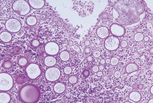


Treatment consists of surgical excision followed by cautery. Injection of amphotericin B at the site of the polyp or topical treatment with dapsone may be efficacious. Relapses occur in up to 10% of the cases.


THE GENUS PNEUMOCYSTIS


Since its discovery in rats by Carlos Chagas in 1909, the taxonomic placement of Pneumocystis has been controversial. It was first described as a trypanosome and was later classified with the protozoa because of similarities in morphology and susceptibility to antiprotozoal drugs. Nucleotide sequence and gene structure data indicate numerous similarities to fungi, including the presence of β-1,3 D-glucan in its cell wall and homologous rRNA and mitochondrial sequences. Despite the fact that Pneumocystis lacks the major fungal sterol, ergosterol, it has been placed in the kingdom Fungi, phylum Ascomycota, class Archiascomycetes, order Pneumocystidales, and family Pneumocystidaceae.


There are several Pneumocystis species, each residing in a specific mammalian host. Initially, the genus contained only one species, P. carinii, but molecular studies have revealed that this is in fact a group of heterogeneous organisms, genetically isolated from each other, that have undergone genetic and functional adaptation to each mammalian host. A nomenclature system that recognizes these differences uses the tripartite “special forms” (formae speciales) designations. The name of each is related to that of the host species from which it originated. Examples of this trinomial nomenclature are P. carinii f. sp. equi, P. carinii f. sp. macacae, and P. carinii f. sp. suis, for horse, macaque and swine strains, respectively. In 1999, Pneumocystis jiroveci was proposed for the human pathogenic strains, whereas P. carinii was reserved for organisms recovered from animals.


Pneumocystis is an opportunistic pathogen that causes severe pneumonia, usually in immunocompromised individuals. Naturally occurring pneumocystosis has been reported in rodents, rabbits, ferrets, mink, horses, dogs, cats, nonhuman primates, goats, and piglets. Disease is found in animals with acquired, inherited, or drug-induced immunodeficiency syndromes, but the exact host immunologic defects that permit proliferation of the organism are not known. In fact, the lack of resistance to P. carinii could be attributed to any defect in synthesis of or response to cytokines or in antigen processing by alveolar macrophages for T-cell presentation. Case fatality rate is high.


Because of the lack of an in vitro propagation system, the life cycle of this organism and the subsequent epidemiology of pneumocystosis have not been defined. It appears that the normal habitat is the lung and the only part of the life cycle that is known is that involving the mammalian lung. The proposed life cycle includes a sexual and an asexual growth phase. The two main developmental stages are the trophozoite and the cyst. Trophozoites are produced during the asexual growth phase and can probably replicate by binary fission. The haploid trophozoites also replicate sexually by conjugation, and produce diploid zygotes. Zygotes undergo meiosis and mitosis, resulting in precyst formation. Differentiation into a mature cyst results in the production of up to eight intracystic bodies that are released when the mature cyst ruptures and develop into trophozoites.


Transmission of infection does not occur between different mammalian species, and results of several studies suggest that humans do not contract pneumocystosis from animals. Animal investigations have documented airborne transmission, but the infectious form has not been identified. Recent findings suggest that immunocompetent hosts may play a significant role in the Pneumocystis life cycle. It has been hypothesized that the main source of P. carinii is individuals with the disease. Accumulating evidence shows that the mammalian host may acquire this organism early in life, in some cases immediately after birth. Transplacental transmission does not occur in the rat model.


Of the common domestic animal species, P. carinii infection has been reported most often in the horse. The majority of cases are in Arabian foals with combined immunodeficiency disease, or in foals that have been treated with immunosuppressive drugs. Infection with Rhodococcus equi may be an immunosuppressive factor. However, several cases have been documented in immunocompetent individuals. Foals are at risk because of the inherent immunodeficiency that occurs between 2 and 4 months of age, as maternal antibodies wane. Clinical signs are mainly limited to the respiratory tract and include cough, dyspnea, and exercise intolerance. There may be a history of poor response to antimicrobial therapy. Gross lesions include firm, meaty lungs with pink and yellow mottling. Lung parenchyma tends to resist being cut and bulges on cut section. Regional lymphadenopathy also may be observed.


Organisms proliferate extracellularly within the lung alveolus. Lung surfactant proteins adhere to the surface of P. carinii and tight adhesions are formed between adjacent organisms and type I alveolar epithelial cells. Diffuse alveolar injury ensues most likely from involvement of the host immune response through costimulation-dependent T-cell–mediated inflammation.


Antemortem diagnosis may be accomplished by careful cytologic examination of fine-needle aspirates of lung biopsy or bronchoalveolar lavage materials. Organisms are small (3-5 μm in diameter) and may exist in low numbers. Wright-Giemsa–type stains best demonstrate trophozoites and intracystic bodies (Figure 49-2). Trophozoites appear as basophilic, dense, oval, or irregular structures having a lobed surface and a single nucleus. Intracystic bodies appear as aggregates of spherical to oval dense basophilic structures against a thick, foamy background. A direct immunofluorescence test can also be applied to bronchoalveolar lavage specimens. Diagnostic immunohistochemical kits are available for use on fixed tissues, and development of polymerase chain reaction (PCR) techniques has given a new alternative for identification of the organism. Serology is an antemortem method to determine exposure to Pneumocystis.
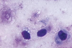


Subacute, interstitial pneumonia with diffuse, alveolar damage, marked macrophage infiltration, and intracellular cysts is observed histologically. Frothy “honeycombed” eosinophilic, intraalveolar material with lymphocytic/plasmacytic interstitial infiltrate suggests P. carinii pneumonia. Fungal stains, such as Gomori methenamine silver, stain the cyst form but not the trophozoite (Figure 49-3). The finding of clusters of nonbudding round to oval to crescent–shaped cysts that appear as “commas” or “parentheses” in alveolar exudates, is confirmatory.
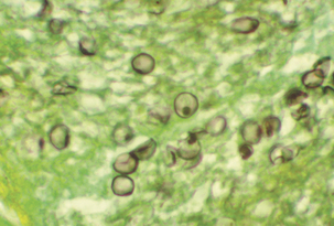

< div class='tao-gold-member'>

Stay updated, free articles. Join our Telegram channel

Jul 18, 2016 | Posted by in PHARMACOLOGY, TOXICOLOGY & THERAPEUTICS | Comments Off on Fungal-Like Agents

Full access? Get Clinical Tree

Get Clinical Tree app for offline access