Chapter 197 Fluoroquinolones
INTRODUCTION
The fluoroquinolone antimicrobial drugs were introduced into clinical medicine approximately 20 years ago. These agents were regarded initially as model antimicrobial agents because of their broad spectrum of activity, favorable pharmacokinetics, and low incidence of toxicity. The fluoroquinolone antimicrobials are entirely synthetic; all possess a common structure containing a 4-quinolone nucleus. Although the more rarely used first-generation fluoroquinolones (nalidixic acid, flumequine) possess a limited spectrum of activity, structural modification of the quinolone nucleus has resulted in an increase in potency and a diversification of spectrum in subsequent generations.
As a class, they are well absorbed after oral and parenteral administration, have a large volume of distribution, and have extended elimination half-lives, allowing for longer dosing intervals. Fluoroquinolones are bactericidal antibiotics at relatively low tissue concentrations and have a favorable margin of safety. Adverse effects in veterinary medicine are associated most frequently with the gastrointestinal (GI) system; however, these agents have been associated with orthopedic, ophthalmologic, neurologic, renal, and cardiac toxicity. This chapter reviews chemical, microbiologic, pharmacokinetic, pharmacodynamic, clinical aspects, and toxicity associated with fluoroquinolone use.
STRUCTURE AND PHYSICAL PROPERTIES
Fluoroquinolones are weak organic acids. They have amphoteric properties as a result of having an acidic group (carboxylic acid) and a basic group (tertiary amine); they are soluble in both alkaline and acidic solutions. All quinolone derivatives in clinical use have a dual ring structure with a nitrogen at position 1, a carbonyl group at position 4, and a carboxyl group attached to the carbon at the 3 position of the first ring (Figure 197-1).1

Figure 197-1 All quinolone derivatives in clinical use have a dual ring structure with a nitrogen at position 1, a carbonyl group at position 4, and a carboxyl group attachedto the carbon at the 3 position of the first ring. The structural modifications to the original dual ring tocreate many of the available quinolone antibiotics are shown in this figure.
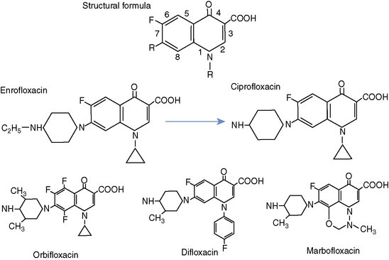
Earlier fluoroquinolones, such as nalidixic acid, did not achieve systemic antibacterial levels. As a result, these agents had limited clinical utility and were suitable only for treating lower urinary tract disease. Fortunately, several structural modifications to the original dual ring have resulted in increased potency, extended spectrum, and enhanced bioavailability. For example, the addition of a fluorine at position 6 led to increased efficacy against both gram-negative and gram-positive bacteria, and substitutions at position 7 result in increased potency and increased antipseudomonal activity. At position 8, addition of a halide, fluorine, or a methoxy group enhances activity against anaerobic bacteria (see Figure 197-1).1 A more extensive discussion of the relationships between structure and activity of the quinolone class is beyond the scope of this chapter.
MECHANISM OF ACTION
Fluoroquinolone antibiotics exert their antimicrobial effect by inhibiting two enzymes of the topoisomerase class: DNA gyrase, or bacterial topoisomerase II, and topoisomerase IV. It is thought that DNA gyrase is the primary quinolone target for gram-negative bacteria, and topoisomerase IV is the target for gram-positive bacteria. For bacterial replication to proceed, individual strands of bacterial DNA must be separated. This results in “supercoiling,” or excessive positive coiling, of DNA strands in front of the replication fork. DNA gyrase is responsible for inducing continuous negative supercoils in the bacterial DNA strand. In addition, DNA gyrase is responsible for removing positive superhelical twists that accumulate ahead of the DNA replication fork through the breakage of both strands of duplex DNA, passage of another segment of DNA through the break, and resealing of the break.1 Both of these actions help to relieve the topologic stress of replication.
DNA gyrase is composed of two subunits (A and B) that must function together for supercoiling to proceed. The A subunit, which is responsible for the strand-cutting function of the gyrase, is the presumed site of action of the quinolones.1 Although inhibition of DNA gyrase leads to functional disturbances that result in rapid death of bacteria, the molecular mechanisms responsible for this bactericidal effect are still not incompletely understood.
Quinolones also inhibit the activity of topoisomerase IV. Topoisomerase IV resolves (decatenate) interlinked (catenated) daughter DNA molecules to facilitate their segregation into their respective daughter cells following replication.1 Inhibition of topoisomerase IV is responsible for the bactericidal effect of the quinolones on gram-positive bacteria.
SPECTRUM
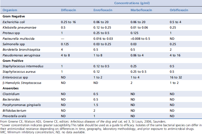
Although the fluoroquinolone antibiotics all possess the same basic chemical structure, agents within the class exhibit variability in their spectrum and potency (Table 197-1). These antibiotics differ from other antibiotics such as penicillins, tetracyclines, and macrolides in that they exhibit a high degree of efficacy at relatively low serum concentrations. In addition, minimal bactericidal concentrations of quinolones are usually within two-fold to four-fold of the minimum inhibitory concentration (MIC) for many of their target organisms.
Table 197-1 Relative Activity of Veterinary Quinolones Against Bacteria Isolated From Animals, as Determined by MIC 90*


Stay updated, free articles. Join our Telegram channel

Full access? Get Clinical Tree

