CHAPTER 13 Field Necropsy of the Horse
For many veterinarians, especially those working in equine and food-animal medicine, the prospect of performing a necropsy in the field, where carcass handling equipment and support personnel are not available, can be a daunting prospect. Although it is common for individuals to undertake a “cosmetic” postmortem examination (i.e., opening the carcass enough to extract the organ system(s) of interest to the case), it is less common for veterinarians to perform a complete necropsy on the animal. In contrast to cosmetic postmortem examinations, a complete necropsy is performed in an effort to systematically examine the entire carcass. This process, though more time consuming and laborious, will greatly enhance the chances of arriving at a diagnosis and provides a more complete understanding of the case.
Whereas there are numerous techniques and means of performing a necropsy, it is very important that the individual performing the procedure develop and use a standard method. As a method is adopted and used, the time required to perform a complete examination will decrease such that it can, and should, become a regular part of the clinical workup of cases. As the practitioner becomes more familiar with the anatomy and appearance of the various body systems after death (and thus able to discern and ignore nonlesions), the process will be expedited.
The necropsy technique that I use and that is outlined herein is based on a technique that is more thoroughly described in The Necropsy Book, published by the C.L. Davis Foundation. In addition to describing and illustrating the necropsy technique, the authors of this book also provide an overview of the most common nonlesions and patterns of a variety of lesions within major organ systems.
PREPARING TO PERFORM THE NECROPSY
Materials Needed to Perform the Field Necropsy
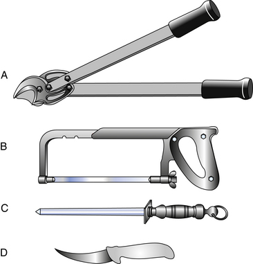
The list of essential tools and materials necessary to perform a field necropsy is relatively short and equipment is easily acquired through a variety of retail establishments that carry landscape supplies and tools (Figure 13-1, Box 13-1). A durable, high-quality necropsy knife (deboning knife) is the one indispensable tool for performing a necropsy; the knife should be purchased from a company that sells knives manufactured for such purposes. It is essential that the person performing the necropsy take the necessary precautions to protect against potential contamination from the carcass by wearing protective clothing, gloves, and eyewear.

Figure 13-1 Essential tools for the field necropsy include rib cutters (A), hand saw (B), knife sharpening steel (C), and knife (D).
Choosing the Site for the Field Necropsy
After you have decided to perform the necropsy, it is important to choose a site that is appropriate with regard to the logistics of carcass removal as well as biosecurity issues. The carcass should be moved to an area that is readily accessible to the equipment that will be used to remove the remains, while at the same time minimizing movement of the remains near paddocks, pastures, and stalls that house other horses on the property. If possible, the necropsy should be performed on a surface that is easily cleaned and disinfected, such as a concrete pad.
The number of people involved with the necropsy should be kept to a minimum and should not involve individuals involved in the daily care of other animals on the property. This will lessen the likelihood of spreading potentially infectious diseases to other horses on the property.
DOCUMENTING THE NECROPSY FINDINGS
Thorough documentation of the necropsy findings should accompany a complete clinical history, and tissue samples should be sent for analysis. Documentation of necropsy findings should be as complete as possible, and findings should be described in accepted medical terminology including use of metric units for measurements.The necropsy report is part of the horse’s medical record and should be written as such.
The essential descriptive features of the necropsy should include (1) location within the organ where the lesion was found, (2) size of lesions, (3) shape of the lesions, (4) color of the lesions, (5) any distinct textural or consistency features, (6) number of such lesions detected or percentage of the organ involved, and (7) description of the contents of a lesion if there is a cavity or space for materials to accumulate. If a digital camera is a available, it is valuable to photograph any areas of interest and to submit these images, along with the case materials, to the diagnostic laboratory.
COLLECTING SAMPLES DURING THE NECROPSY
As the necropsy is performed and the various organ systems are examined, a small sample of each tissue (maximum thickness, 0.5 cm) should be immersed in a container containing 10% neutral buffered formalin for histologic evaluation. For optimal, rapid fixation, there should be a 10:1 ratio between the volume of formalin and the tissues for fixation. The brain and the eye are the only organs that should be fixed in their entirety. The brain should be fixed separately, and it is advisable to inject formalin into the vitreous chamber of the eyes before immersion.
It is also advisable to collect a complete survey of organs from the horse for histopathologic evaluation, although not all tissues will necessarily be processed and analyzed. It is better to submit too many tissue samples than not to have sampled an organ that would have been valuable in understanding the case and making a diagnosis. The decision as to which organs to process for histologic assessment can be made by the pathologist after reviewing the case history and consulting with the submitting veterinarian.
Coincident with collecting tissues for histologic evaluation, it is advisable to collect fresh, unfixed tissues for additional ancillary testing (e.g., microbiology, toxicology). These samples should be placed in sealable plastic bags and sent on ice to the diagnostic laboratory for any additional necessary testing.

Full access? Get Clinical Tree

