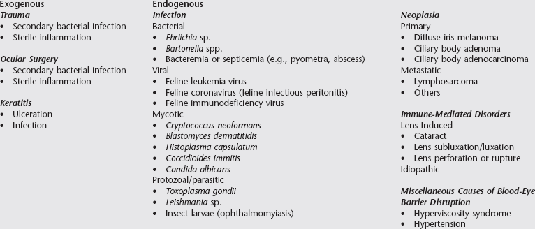
Feline Uveitis

Chapter 250


Feline Uveitis


Uveitis is a painful, vision-threatening disease common in cats. Previous reports suggest that 40% to 70% of feline uveitis cases are associated with a systemic illness. A thorough workup to eliminate infectious causes is imperative because treatment of idiopathic uveitis is directed toward immunosuppression. Owners should be prepared for a potentially expensive diagnostic workup in which a definitive diagnosis is not identified. Some causes of uveitis in cats are different from those identified in the dog (see Chapter 249).


The uveal tract is comprised of the iris, ciliary body, and choroid. Anterior uveitis or iridocyclitis is inflammation of the iris and ciliary body. Posterior uveitis is more commonly referred to as chorioretinitis since the choroid and retina are affected concurrently in many cases. Anterior uveitis and chorioretinitis can occur separately or together.


Clinical Manifestations


Intraocular inflammation is initiated by tissue injury from trauma, infectious agents, or other antigen challenge. The tissue factors released from the damaged tissue and microorganisms result in vasodilation and changes in the vascular permeability of the intraocular vasculature. Inflammatory mediators cause leukocyte activation and migration. With moderate or severe anterior uveitis, increases in protein concentrations and inflammatory cells numbers lead to opacity of the normally transparent aqueous humor, also called aqueous flare. Hyphema, fibrin clots, and keratic precipitates (inflammatory cell and protein aggregates on the corneal endothelium) also may be observed. Conjunctival and scleral injection, photophobia, blepharospasm, enophthalmos, and epiphora are common with anterior uveitis but often are not present with chorioretinitis unless anterior uveitis also is present. Other ocular signs of anterior uveitis are corneal edema, miosis, edema and color changes in the iris, anterior and posterior synechia, and iris bombé. Decreased intraocular pressure (IOP) frequently is observed as a consequence of decreased aqueous humor formation. Normal feline IOP is reported as approximately 10 to 20 mm Hg. Ocular hypotony should resolve as the intraocular inflammation is controlled. Normal IOP in an actively inflamed eye raises the suspicion of concurrent glaucoma. Trends in IOP should be monitored closely throughout treatment.


Inflammatory cells sometimes accumulate in the anterior vitreous, obscuring the retina from direct visualization. This is referred to as pars planitis or intermediate uveitis. In the choroid and retina, exudates of protein and inflammatory cells accumulate within and beneath the retina and cause retinal and subretinal edema, hemorrhage, and retinal detachment. Since the retina and subretinal space are immediately anterior to the tapetum, tapetal reflectivity is decreased or obscured by areas of active chorioretinitis.


Diagnosis


Causes of feline uveitis generally are subdivided into the categories of trauma, infection, immune-mediated disease, and neoplasia (Box 250-1). Some causes can be identified by history (trauma) or ocular examination (trauma, lens disorders, neoplasia). Correlating infectious disease with uveitis is more challenging. Infectious causes generally cannot be differentiated by ocular findings alone, so the history, physical examination findings, and results of a complete blood count, urinalysis, and serum biochemical panel should be used to guide further diagnostic testing such as serologic studies, aqueous or vitreous humor analysis, radiography, ocular ultrasonography, and histopathologic evaluation. Ocular fluids can be used for cytologic analysis, culture and sensitivity testing, polymerase chain reaction (PCR) assay, and determination of antibody content.


Infectious Diseases Associated with Feline Uveitis


Toxoplasmosis


Infection with Toxoplasma gondii occurs primarily by ingestion of tissue cysts in prey animals. Chorioretinitis is the most common ocular manifestation of toxoplasmosis in systemically ill cats; however, the majority of infected cats do not become systemically ill. Cats with positive results on tests for T. gondii infection often exhibit anterior uveitis but are otherwise clinically normal. Evidence suggests that T. gondii–related anterior uveitis is immune mediated. Definitive diagnosis of ocular toxoplasmosis is difficult due to the high percentage of cats with serum titers showing positivity for T. gondii. The presence of serum immunoglobulin M, a rising serum immunoglobulin G level, and antibody production in the aqueous humor all support a diagnosis of ocular toxoplasmosis.


All sick cats that test positive for T. gondii should be treated with anti-Toxoplasma drugs. T. gondii-positive cat with uveitis, but no other clinical signs of infection by T. gondii, should be treated with anti-Toxoplasma drugs when other causes have been ruled out, especially if the uveitis responds poorly to antiinflammatory therapy alone. Clindamycin hydrochloride is recommended at 10 to 25 mg/kg q12h for 14 to 21 days.


Feline Infectious Peritonitis


Ocular disease (anterior and posterior uveitis) associated with feline infectious peritonitis (FIP) is most common in the dry, noneffusive form of FIP and usually is accompanied by signs of systemic disease. Rarely ocular signs may be the sole presenting complaint. FIP typically occurs in cats younger than 3 years of age. Currently available tests cannot distinguish antibodies against feline enteric coronavirus from those against FIP-inducing coronavirus; however, in a young cat with ocular disease, the findings of a coronavirus antibody titer of more than 1 : 1600, lymphopenia, and hypergammaglobulinemia are highly suggestive of FIP infection. Once clinical signs occur, the prognosis for FIP is grave. Cats with less severe disease may live for several months with therapy.


Immunosuppressive and antiinflammatory drugs may be used for palliative therapy. Several protocols have been described, but there is no consensus on optimal treatment of systemic disease. Ocular inflammation should be treated with topical and systemic glucocorticoids and with cycloplegics, depending on the location and severity of the inflammation (see treatment section).

< div class='tao-gold-member'>

Stay updated, free articles. Join our Telegram channel

Jul 18, 2016 | Posted by in PHARMACOLOGY, TOXICOLOGY & THERAPEUTICS | Comments Off on Feline Uveitis

Full access? Get Clinical Tree

Get Clinical Tree app for offline access