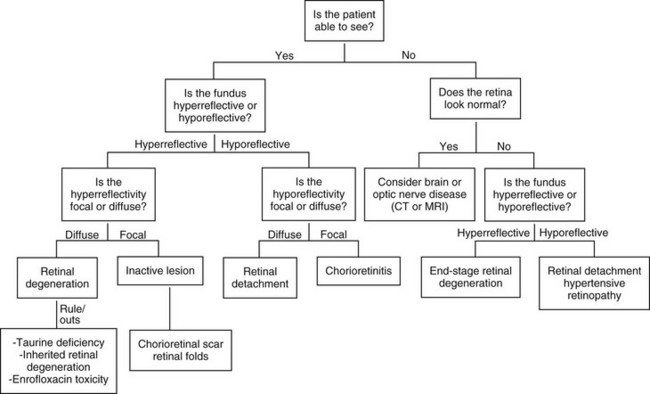
Feline Retinopathies

Chapter 255


Feline Retinopathies


Examination of the feline retina is an important part of a thorough systemic evaluation. Any cat with a pupillary abnormality, a presenting complaint of vision loss, a systemic illness of unknown origin, or a cardiac abnormality should undergo a complete retinal examination. Proper retinal evaluation can help the practitioner to focus the diagnostic search as well as monitor response to therapy. This chapter reviews the most common causes of retinal lesions in the feline and presents a diagnostic approach to retinal changes. Figure 255-1 provides a diagnostic algorithm for feline retinal disease.


The biggest obstacle to understanding retinal disease is the difficulty inherent in fundic examination. The fundus is defined as the visible structures in the posterior half of the eye and includes five overlapping layers of tissue with varying degrees of transparency. The fundus is composed of the sclera, the choroid, the tapetum, the retinal pigmented epithelium, and the neurovascular retina. The posterior blood-eye barrier is formed by the nonfenestrated capillaries of the neurovascular retina and the tight junctions between retinal pigmented epithelium cells, and acts to prevent substances in the bloodstream from entering the eye. Pathologic conditions result in a breakdown of this barrier, which can allow offending substances, including inflammatory cells, infectious organisms, or blood, to collect within and under the retina. These alterations in the layers of the fundus result in the characteristic changes in the appearance of the retina.


The first key to effective fundic evaluation is an understanding of the normal retinal appearance. Performing routine retinal examinations and using an ophthalmic atlas will help to establish a thorough recognition of normal variation. When retinal disease is detected, it is first important to differentiate active from inactive disease. When cells or fluid sit between the retina and the tapetum the tapetal reflection is blocked, which leads to a murky or blurry image. Thus active retinal lesions often are hyporeflective and fuzzy with irregular borders. Atrophy of the retina results in vascular attenuation and retinal thinning, which allows greater tapetal reflection. Thus inactive lesions are hyperreflective with sharp margins. Inactive lesions also may be associated with pigment clumping, leading to foci of hyperpigmentation within or adjacent to a hyperreflective lesion.


Hypertensive Retinopathy


Hypertensive retinopathy is perhaps the most common and clinically important retinopathy in the cat. It should be considered in all cases of acute blindness in older cats. Hypertensive retinopathy has been used as a broad term to include both hypertensive retinopathy and choroidopathy. The retinal arteries autoregulate in response to increased systemic blood pressure. This results in vasoconstriction, which in turn leads to hypertrophy of the smooth muscle layer of the arteriole and ultimately to focal necrosis and rupture of the vessel. These focal ruptures produce multifocal areas of retinal edema or hemorrhage. As more vessel damage occurs, serum and blood continue to leak, which leads to complete retinal detachment and blindness. Additionally, the choroid does not autoregulate, and as the blood pressure increases, there is a substantial degree of choriocapillaris serum leakage resulting in serum collection beneath the retina and exudative retinal detachment.


The incidence of ocular signs in hypertensive cats is about 40% to 60%, and ocular signs are the most common form of target organ dysfunction associated with systemic hypertension. Hypertensive retinopathy is identified most often in cats older than 10 years of age with systolic blood pressures greater than 168 mm Hg when measured by an oscillometric technique (Sansom et al, 2004). The presence of ocular changes is an indication to start antihypertensive therapy even if blood pressure measurements do not consistently meet the criteria for hypertension.


Diagnosis


Clinical signs of hypertensive retinopathy can be unilateral but are typically bilateral. Presenting complaints include blindness, vision loss, or progressively dilated pupils. Hypertensive retinopathy also may have a subclinical presentation, noted only during routine examination of the fundus or during examination of cats with high blood pressure or those evaluated for a gallop sound or heart murmur. Serous retinal detachment often can be diagnosed with a penlight from arm’s distance. Retroillumination of the eye is achieved by holding a light at arm’s length from the patient. A normal eye should show bright yellow-green tapetal reflection in the pupil, although a blue eye may have a red pupil due to a lack of tapetum. Retinal detachment results in a dampening of that reflection caused by the presence of fluid between the retina and the tapetum. The proximity of the retina to the lens also sometimes allows the retinal vessels to be seen directly through the pupil. Retinal examination is difficult with retinal detachment, and the entire fundus may seem blurry. Focal areas of detachment are blurry and hyporeflective and are sometimes associated with hemorrhage beneath, within, or above the retina. Hyphema or vitreal hemorrhage also may be observed.


Treatment and Prognosis


Treatment of hypertensive retinopathy is focused solely on control of the systemic hypertension. No topical medications are indicated to treat the ocular component. Amlodipine has been demonstrated to be particularly effective in the resolution of systemic hypertension and hypertensive retinopathy (Maggio et al, 2000). Current recommendations for the treatment of systemic hypertension can be found in Chapter 169, but cats with hypertensive retinopathy should be evaluated for underlying systemic disease, including renal failure, hyperthyroidism, and the more uncommon condition of hyperaldosteronism (Conn’s syndrome).


The prognosis for vision varies depending on the duration of the retinal detachment, with experimental models showing a return to function if the retina has been detached for 2 weeks or less. Unfortunately, because of cats’ ability to adjust to vision loss, the retina typically is detached long before the patient is seen by a clinician. Although resolution of the hypertension usually results in reattachment of the retina, continued retinal degeneration is likely to occur. Thus some cats who are visual at presentation or regain vision immediately after treatment may continue to go blind despite adequate control of blood pressure.

< div class='tao-gold-member'>

Stay updated, free articles. Join our Telegram channel

Jul 18, 2016 | Posted by in PHARMACOLOGY, TOXICOLOGY & THERAPEUTICS | Comments Off on Feline Retinopathies

Full access? Get Clinical Tree

Get Clinical Tree app for offline access