Feline mycobacterial disease

47 Feline mycobacterial disease


INTRODUCTION


Most cutaneous and subcutaneous abscesses, cellulitis and wounds seen in cats are due to fights, and are infected by both aerobic and anaerobic bacteria. Pasteurella multocida, B-haemolytic streptococci, Actinomyces spp., Bacteroides spp. and Fusobacterium spp. are commonly isolated from abscesses. In most cases, surgical drainage and routine antibacterial therapy resolves the infection.


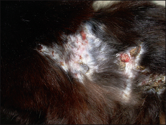
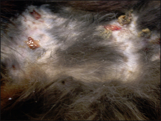
Non-healing draining wounds and nodules, however, although uncommon, can be a diagnostic and therapeutic challenge. They can be caused by infectious organisms (bacterial, fungal, viral and protozoal organisms), foreign body reactions, sterile panniculitis and neoplasia (Table 47.1). Concurrent infections with FIV, FeLV or corona virus and the use of immunosuppressive drugs may be underlying causes of infection. Identification of the infectious organism is important, as some may have zoonotic implications and others may be life threatening. Cats presented with non-healing abscesses, non-healing wounds, nodules, draining tracts and cellulitis require extensive investigation and treatment.


Table 47.1 Causes for cutaneous nodules and draining tracts in cats


Bacteria Non-acid-fast organisms Actinobacillus spp., Arcanobacterium pyogenes (previously known as Actinomyces pyogenes), Rhodococcus equi, Staphylococcus spp., Streptococcus spp., Pseudomonas spp., Proteus spp.
Acid-fast organisms Norcardia asteroides, Mycobacterium lepraemurium, M. tuberculosis, M. microti, M. chelonei, M. fortuitum, M. phlei, M. thermoresistible, M. smegmatis
Fungi Subcutaneous mycoses M. canis, Rhizomucor, Mortierella, Fusarium, Paecilomyces, Alternaria, Cladosporium, Exophiala, Moniliella, Curvularia, Madurella, Pythium insidiosum, Sporothrix schenckii
Systemic mycoses Cryptococcus neoformans, Sporothrix schenckii, Histoplasma capsulatum, Blastomyces dermatidis, Coccidioides immitis
Parasitic or protozoal   Leishmania spp., cutaneous habronemiasis
Viral   Cowpox virus, calcivirus, herpes virus
Non-infectious causes   Foreign body reactions, eosinophilic granulomas, sterile nodular panniculitis, xanthamatosis, cutaneous histiocytosis and neoplasia

After Patel A (2002): Pyogranulomatous skin disease and cellulitis in a cat caused by Rhodococcus equi. J Small Anim Pract 43, 129–132, with permission of Blackwell Publishing.


One of the causes of persistent non-healing wounds, nodules and draining sinus tracts in cats is infection with rapidly growing mycobacteria (previously known as opportunistic or atypical mycobacteria). The report describes the investigation and treatment of such a case in which there was also a concurrent Nocardia infection.


CASE HISTORY


The various forms of feline mycobacterial disease are poorly responsive to short courses of antibiosis used for the treatment of routine cat-bite abscesses. There may be a history of a fight or the individual may have a history of hunting small rodents. Some individuals may have had surgery in an attempt to remove the diseased tissue, and this sometimes results in wound breakdown and subsequent failure to heal. The cat may have a history of anorexia, weight loss and pain.


In this case the relevant history was:


CLINICAL EXAMINATION


In general, cats with infections caused by the rapidly growing mycobacteria (RGM) have few signs of systemic illness, although more extensive disease may result in pyrexia, reluctance to move, peripheral lymphadenopathy and other constitutional signs. Typically, fight wounds are the cause of RGM infections and lesions tend to start in areas subject to biting and scratching such as the dorsum, flanks and inguinal fat pad. Early on in the clinical course, the lesions resemble catfight abscesses, but they generally do not have the putrid malodour associated with them. As lesions progress, the affected areas become alopecic with numerous discharging punctuate sinus tracts. The surrounding subcutis may be thickened and the overlying skin may become adherent to the underlying tissue. Some areas may have ulcers of varying sizes and necrotic skin. Hypercalcaemia has been reported in some cats.


A clinical examination revealed:


Stay updated, free articles. Join our Telegram channel

Sep 3, 2016 | Posted by in SMALL ANIMAL | Comments Off on Feline mycobacterial disease

Full access? Get Clinical Tree

Get Clinical Tree app for offline access