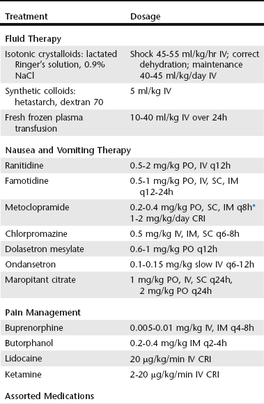
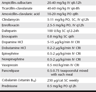
Feline Exocrine Pancreatic Disorders

Chapter 138


Feline Exocrine Pancreatic Disorders


With advances in serology and diagnostic imaging, disorders of the exocrine pancreas in cats are detected more frequently. Pancreatitis has been recognized as a relatively common disorder causing significant morbidity and, infrequently, mortality. Less-common disorders of the exocrine pancreas include exocrine pancreatic insufficiency (EPI), pancreatic cancers (adenocarcinoma, lymphoma), pancreatic pseudocyst, abscess, parasites, and nodular hyperplasia. This chapter focuses on diagnostic confirmation and therapeutic management of mild to moderate and severe pancreatitis, complications of pancreatitis, and areas of uncertainty in the management of pancreatitis, including the use of antibiotics and corticosteroids.


Pancreatitis is characterized histologically as acute (primarily neutrophilic), chronic (primarily lymphocytic or lymphoplasmacytic with fibrosis), or acute component with chronic disease (both neutrophilic and lymphocytic). The prevalence of cats with clinical pancreatitis is unknown; pancreatic inflammation is a relatively common necropsy finding (very mild to severe lesions in 67% of cats evaluated). In contrast to dogs, the two most frequent clinical signs of pancreatitis in cats are lethargy (88% to 100%) and anorexia (95% to 97%). Less commonly, cats display weight loss (47%), vomiting (35% to 52%), and infrequently diarrhea (11% to 38%), labored breathing (20%), or polyuria/polydipsia (20%). Common physical examination findings include dehydration (92%), hypothermia (68%), and less frequently icterus (37%), palpable abdominal mass (23%), apparent abdominal pain (15%), and rarely fever (7%). Unlike dogs, many cats with pancreatitis have a normal complete blood count (CBC) with abnormalities such as neutrophilia (46%), nonregenerative anemia (38%), hemoconcentration (17%), and leukopenia (15%). Hypercholesterolemia (72%) frequently is noted on a biochemical panel with less common findings of hyperbilirubinemia (58%), increased liver enzymes (alanine transaminase [ALT] 57%, alkaline phosphatase [ALP] 49%), hypokalemia (56%), hyperglycemia (45%), and hypoalbuminemia (36%). Hypoglycemia and ionized hypocalcemia (65%) are a frequent finding in cats with severe, necrotizing pancreatitis, and these patients require more aggressive supportive care with a guarded prognosis. Despite these nonlocalizing clinical signs, physical examination findings and laboratory abnormalities, the frequency of the antemortem diagnosis of pancreatitis in cats has increased with advances in pancreatic serology and imaging.


Diagnosis of Feline Pancreatitis


Accurate diagnosis of pancreatitis involves a combination of appropriate clinical signs and physical examination findings, laboratory abnormalities including elevated pancreatic-specific lipase, and ultrasonographic changes. Pancreatic cytology or histopathology infrequently is needed for confirmation but can be beneficial in the treatment of pancreatitis, in particular if severe or with complications. Multiple generations of pancreatic enzyme serology testing have resulted in improvements in the diagnostic accuracy for feline pancreatitis. Serum amylase, lipase, and trypsin-like immunoreactivity (TLI) concentrations are of limited diagnostic value for pancreatitis; however, TLI is the preferred diagnostic test for EPI. Although not evaluated by a research study, higher ascites lipase concentrations compared with serum lipase concentrations have been associated with pancreatitis, in the author’s experience. The current assay (Spec fPL) measures pancreatic-specific lipase by a monoclonal antibody sandwich enzyme-linked immunosorbent assay (ELISA) with a moderate sensitivity and specificity, 79.4% and 79.7%, respectively. Transabdominal pancreatic ultrasound is a moderately sensitive test (73%) for pancreatitis in cats; however, it has a variable specificity (24% to 67%) and depends on available equipment and sonographer skill level. However, unlike serology, ultrasound imaging permits detection of nonpancreatic concurrent disorders, screening for causes of pancreatitis, and ultrasound-guided fine-needle aspiration cytology of the pancreas and peripancreatic fluid accumulations. Using pancreatic-specific lipase and ultrasound imaging in the diagnosis of pancreatitis provides the highest positive predicative value, 69% and 80%, respectively, and negative predictive value, 87% and 57%, respectively.


Ultrasonographic changes associated with pancreatitis include pancreatomegaly, hypoechoic pancreatic parenchyma, hyperechoic peripancreatic fat or mesentery, dilated pancreatic or bile duct(s), dilation of the gallbladder, thickened gastric wall, and corrugated, thickened duodenal wall and peripancreatic fluid accumulation (ascites). The minimum requirements for the ultrasonographic diagnosis of pancreatitis in cats have not been determined. Certain abnormalities, including increasing pancreatic duct size and, potentially, pancreatic parenchyma hyperechogenicity, may occur in older cats without pancreatitis. Pancreatic computed tomography (CT), the standard diagnostic test for certain types of pancreatitis in humans, has poor specificity and sensitivity in cats.


Treatment of Mild to Moderate Feline Pancreatitis


Clinical signs of pancreatitis range in severity from mild and self-limiting to severe, life-threatening disease with an acute or chronic presentation. In contrast to dogs, the cause of pancreatitis in cats is not associated with dietary fat intake, obesity, or drugs. Specific causes include cancer, infections (viral [feline infectious peritonitis (FIP), feline immunodeficiency virus (FIV), calicivirus], Toxoplasma gondii, Amphimerus pseudofelineus), and organophosphates. In addition, up to two thirds of cats with pancreatitis have concurrent disorders, including hepatic lipidosis, cholangitis, obstructive jaundice, inflammatory bowel disease, diabetes mellitus, interstitial nephritis, and pleural effusion. It is important to consider these concurrent disorders with persistent or progressive clinical signs despite therapy.


The core therapy for pancreatitis without complications is supportive and symptomatic. Intravenous fluid is used to restore circulating blood volume, antiemetic medications are used to control nausea and vomiting, and pain relief is provided as needed (Table 138-1). Although less frequently detected than in dogs, potentially because cats have less definitive indicators of pain, abdominal pain can contribute to persistent inappetence, and these cats improve with therapy. The author uses buprenorphine or butorphanol as first-line pain relief medication in cats with mild to moderate pancreatitis. Nutritional support is an important component of therapy. Restriction of oral food and water intake in a nonvomiting cat is not considered necessary; rather, oral intake of food is encouraged. Fasting cats with pancreatitis can lead to development of malnutrition, intestinal atrophy, bacterial translocation, and hepatic lipidosis. With mild to moderate pancreatitis, appetite stimulants are often effective to encourage voluntary intake of food. However, with persistent inappetence for more than 3 to 4 days, placement of an esophageal or gastric feeding tube (percutaneous endoscopic gastrostomy [PEG]) should be considered. The ideal dietary composition to feed cats with pancreatitis has not been determined; however, in contrast to dogs, marked fat restriction is likely not necessary. Easily digested, enteral diets currently are recommended. Furthermore, for cats with mild to moderate pancreatitis, the author does not treat with antibiotics or corticosteroids (see section on Areas of Uncertainty later in the chapter).


< div class='tao-gold-member'>

Stay updated, free articles. Join our Telegram channel

Jul 18, 2016 | Posted by in PHARMACOLOGY, TOXICOLOGY & THERAPEUTICS | Comments Off on Feline Exocrine Pancreatic Disorders

Full access? Get Clinical Tree

Get Clinical Tree app for offline access