36 Feline eosinophilic plaque
CASE HISTORY
In this case the relevant history was:
• The first sign of disease was 2 years previously and consisted of pruritic lesions over the groin and medial thighs. These lesions resolved with a single injection of a depot glucocorticoid, methylprednisolone acetate.
• Since then there had been further similar episodes, which also responded to depot glucocorticoid injections. However, the most recent episode had failed to respond despite multiple injections given at a higher dosage.
• The cat received routine thorough flea control consisting of monthly applications of fipronil spot-on and the house environment had also been treated with a permethrin and pyriproxyfen spray twice in the last 3 months.
CLINICAL EXAMINATION
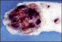
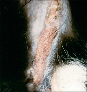
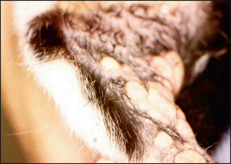
Other variants of the eosinophilic granuloma complex may be found on the same individual, e.g. ‘linear granuloma’, which is seen as a raised chord-like linear plaque, with a yellowish surface, on the caudal aspect of the thighs (Fig. 36.2). Indolent (rodent) ulcer may also be seen as an area of ulceration on the upper lip. Some individuals may also have lesions on the hard or soft palates.
In this case the clinical examination revealed:
CASE WORK-UP
In the first instance, the following investigations were carried out:
• An impression smear of the lesion, stained with modified Wright’s stain, revealed a mixed inflammatory cell population, mainly of eosinophils and neutrophils. Extracellular and intracellular coccoid bacteria were also present (Fig. 36.4).
• A swab sample was cultured, which revealed coagulase-negative staphylococci (speciation was not carried out on the sample).
• Blood samples for routine haematological and biochemical parameters, which were within laboratory reference ranges.
• Skin scrapes, coat brushings and fungal culture from toothbrush combing, which were all negative for ectoparasites and dermatophytes.
• Three skin samples were taken by punch biopsy for histology, one from the centre of the lesion, one from the edge of the lesions and one from the surrounding skin. (The diagnosis of eosinophilic plaque may be made on clinical appearance and cytological examination alone but histopathological examination is indicated if there is any doubt about the diagnosis and in this case ruled out a number of the other differentials.)
Only gold members can continue reading. Log In or Register to continue
You may also need

Full access? Get Clinical Tree

