Chapter 11 Failure of Epithelial Migration: Ceruminoliths
The ear canal requires a clearance mechanism to remove the accumulation of dead cells, trapped foreign debris, and wax. Without a physiologic method of debris removal, large accumulations of material would remain in the ear canal.
Epithelial Migration
Fortunately, the ordered growth of the ear canal epithelium facilitates a clearing mechanism termed epithelial migration. Simply stated, the epithelium in the ear canal grows outward from the tympanic membrane toward the opening of the external ear canal. As the surface epithelial cells move, they carry along any debris on top of them. This physiologic epithelial movement process may be demonstrated by placing India ink on the eardrum and observing its dispersal along the ear canal over several weeks (Figure 11-1). The rate of epithelial movement is slow, and in older animals and people the rate becomes even slower. When the rate slows to the point of allowing accumulation of debris, the term failure of epithelial migration applies (Figure 11-2). When this condition causes accumulation of substances within the ear canal, ear flushes and ceruminolytic agents play important roles in its management.
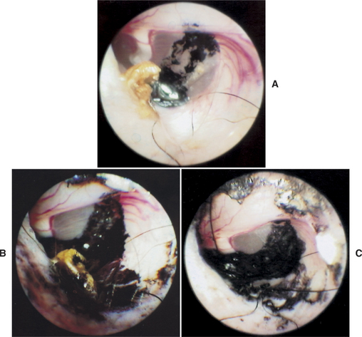
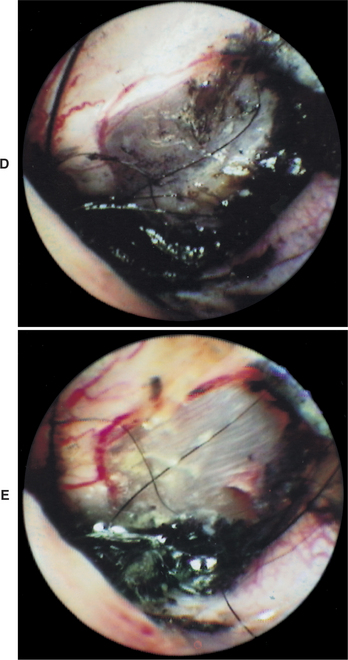
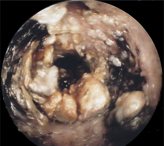
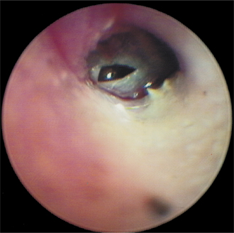
Figure 11-2 Accumulation of ceruminous exudates in the ear. Epithelial migration is not able to remove the waxy matter.
Besides playing a role in protecting the ear canal as a mechanical barrier to environmental substances, the keratinocytes have been shown to possess immune functions. Interleukin-1 (IL-1) is stored in keratinocytes. When these cells are damaged, IL-1 is released, stimulating other cells to release more IL-1, and a cascade of immunologic events results in the migration of granulocytes, monocytes, and macrophages into the site of damage. When an area of the ear canal has been denuded by trauma or ulcer formation, loss of this protective immune mechanism allows unchecked bacterial colonization, favoring the development of otitis externa.
Failure of Epithelial Migration
Damage to the germinal epithelium of the eardrum from infection or ear mite infestation results in damage to the keratinocytes on the surface of the tympanic membrane (Figure 11-3). During the healing process, fibrosis may cause permanent changes to the tympanic membrane. Normal epithelial migration may be disrupted. Failure of epithelial migration may result in the accumulation of flakes of skin in the ear canal. More often, however, wax and keratin accumulate at the base of the eardrum and form either soft wax plugs (Figure 11-4) or large, hard concretions called ceruminoliths (Figure 11-5). To find them when examining the ear canals with an otoscope, the veterinarian should follow the bend in the ear cartilage and look in the horizontal canal toward the eardrum. Ceruminoliths are found attached to the eardrum lying along the floor of the horizontal ear canal.
Stay updated, free articles. Join our Telegram channel

Full access? Get Clinical Tree


