Chapter 1 Anatomy of the Canine and Feline Ear
The basic anatomical components of the dog and cat ear are as follows:
Structure of the External Ear
The external ear is composed of three elastic cartilages: annular, scutiform, and auricular (Figure 1-1). The annular and auricular cartilages form the external ear canal, and the auricular cartilage expands to form the pinna. The scutiform cartilage lies medial to the auricular cartilage within the auricular muscles that attach to the head (Figure 1-2).
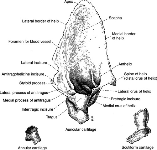
Figure 1-1 Cartilages of the right external ear.
(From Evans HE, ed: Miller’s Anatomy of the dog, ed 3, Philadelphia, 1993, WB Saunders.)
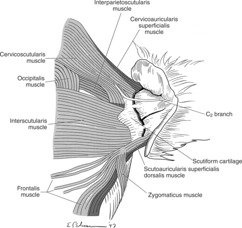
Figure 1-2 Location of the scutiform cartilage in relation to some of the external ear muscles in the dog.
Pinna, or Auricle
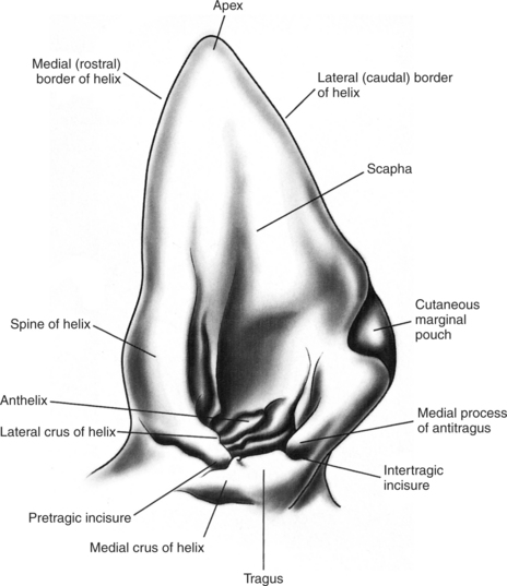
The leaf-shaped pinna of the external ear is broad with medial (rostral) and lateral (caudal) margins. The caudal margin of the pinna exhibits a cutaneous pouch called the marginal pouch (Figure 1-3). This pouch has no obvious function. The skin on the concave surface of the pinna is very tightly connected to the underlying auricular cartilage, accentuating all the auricular prominences (see Figure 1-3). The skin covering the auricular cartilage may show breed-specific pigmentation. The shape and size of the external ear vary greatly among different breeds of dogs, mainly owing to the auricular cartilage that forms the skeleton of the pinna. It is the largest cartilage of the external ear. The broad auricular cartilage has numerous holes (see Figure 1-1), which are traversed by branches arising from the caudal auricular artery.
The auricular cartilage is broad dorsally and funnels to a narrow tubelike structure, the tubus auris, which fits around the annular cartilage ring. The parotid salivary gland occupies the base of the external ear, partially surrounding the tubus auris (Figures 1-4 and 1-5). The tubus auris encloses the vertical part of the external canal and, together with the tragal, antitragal, and antihelicene borders, forms the external acoustic meatus (see Figure 1-3).
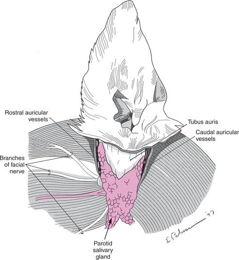

Figure 1-5 Structure of the ear canal.
(Redrawn from Bojrab MJ: Current techniques in small animal surgery I, Philadelphia, 1975, Lea & Febiger.)
Annular Cartilage
The annular cartilage is part of the external ear canal. The pinna, formed by the cone-shaped auricular cartilage, articulates with the annular cartilage. The annular cartilage is a ring-shaped structure attached to the bony orbit of the external acoustic meatus of the temporal bone. A tubular cartilage piece, the annular cartilage surrounds the osseous external acoustic meatus (see Figure 1-7). It is attached to the bony rim of the external acoustic meatus by fibrous tissue that permits some degree of movement of the external ear. The annular cartilage encloses the horizontal part of the external ear canal.
Scutiform Cartilage
The scutiform cartilage is an L-shaped structure located over the temporalis muscle. It does not contribute to the formation of the external ear or its canal. The scutiform cartilage is attached to the midline raphe of the head and neck by numerous muscles (see Figure 1-2). Muscles also extend from the scutiform cartilage to the auricular cartilage. The scutiform cartilage functions like a fulcrum, providing for efficient movement of the auricle. It can be considered to function like a sesamoid cartilage. It lies over a fat cushion (corpus adiposum auriculae) on the dorsal surface of the temporalis muscle.
Structure of the Ear Canal
The external ear canal in the dog is 5 to 10 cm long and 4 to 5 mm wide (see Figure 1-5). The ear canal consists of an initial vertical part, which may extend an inch. The vertical canal runs ventrally and slightly rostrally before bending to a shorter horizontal canal that runs medially and forms the horizontal part of external ear canal. Because the external ear is elastic, the ear canal can be straightened enough to permit otoscopic examination.
Nerves of the External Ear
The facial nerve is related to the ventral surface of the annular cartilage, close to the osseous external acoustic meatus. The facial nerve provides substantial sensory innervation to the concave surface of the scapha and part of the cavum conchae via the rostral, middle, and caudal internal auricular branches (Figure 1-6). Most of the vertical along with part of the horizontal ear canal lining is supplied by the lateral internal auricular branch of the facial nerve, which may contain predominantly vagal fibers.
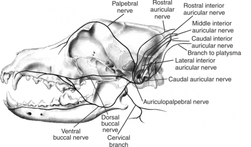
Communication between the facial nerve and the vagal nerve takes place as the facial nerve exits the stylomastoid foramen (see Figure 1-11). It is believed that these vagal branches are given off as the lateral internal auricular branch to the skin of the external ear canal. Reflex gastric vomiting may be triggered if the sensory endings of the vagus nerve are stimulated by mild ear canal irritation.
Stay updated, free articles. Join our Telegram channel

Full access? Get Clinical Tree


