Eyelids – introduction

3 Eyelids – introduction


An understanding of the basic anatomy and physiology of the eyelids is important since so many common ophthalmic problems are encountered in this area, especially in dogs, but less frequently in cats and rabbits as well. The tremendous variation in conformation exacerbated by selective breeding for specific appearance has contributed to the numerous pathological conditions in the area. These can be primarily ocular, such as entropion (rolling in of the eyelids) or distichiasis (extra eyelashes along the eyelid margin) or can be secondary to skin diseases or immune-mediated conditions for example.


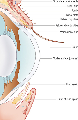
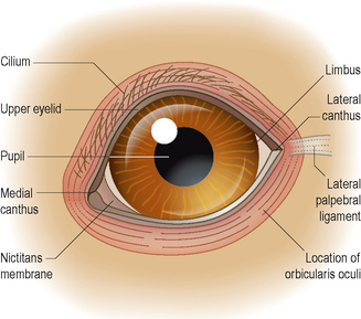
The eyelids are dorsal and ventral folds of skin lined with palpebral conjunctiva and they form the palpebral fissure in which the globe is situated (Figure 3.1). The normal outline of the palpebral fissure is almond shaped (Figure 3.2) but many different variations occur with pedigree animals. The skin should be only loosely adherent to underlying structures but again this can vary from being excessively loose (e.g. in the Saint Bernard) to being very tight (e.g. the miniature poodle). The upper eyelid is more mobile than the lower in most species. A fibrous layer for support, the tarsal plate, is present within the eyelids. This is less well developed in dogs than cats.


In addition to providing support, the eyelid folds also contain both smooth and striated muscles and a good vascular and nerve supply. The most important muscle is the orbicularis oculi. The orbicularis oculi is mainly involved with eyelid closure and acts as a sphincter. It is located immediately below the skin and is innervated by the palpebral branch of the facial nerve (cranial nerve VII). Eyelid sensation is provided by branches of the trigeminal nerve (cranial nerve V). The usual rapid action of this muscle is important for globe protection and in the distribution of the tear film. Ocular pain can result in severe blepharospasm as the orbicularis oculi remains tightly contracted which can lead to further ocular pain with entropion and ulceration.


The normal position of the eyelids is resting against the ocular surface, and they slide over it during blinking. Many breeds of dogs and cats have poor lid–globe apposition and this results in ocular problems such as entropion or ectropion.


Palpebral ligaments are present medially and laterally and these fibrous bands stabilize the lids and help to maintain the shape of the palpebral fissure. The medial canthal ligament is well developed and having originated from the medial fibres of the orbicularis oculi muscle it inserts on the orbital rim of the lacrimal bone. In most dogs the lateral canthal ligament is poorly developed and really consists of lateral fascial attachments only. However, in some breeds, particularly those with broad skulls and relatively deep-set eyes such as the Rottweiler and mastiff types, the lateral canthal ligament is more defined but misdirected medially, resulting in some lateral canthal involution. This needs to be addressed when considering entropion surgery in these breeds and is mentioned in the case on complicated entropion later in this section.


Along the free margin of the lids (especially the upper lid) are a row of cilia (not true eyelashes). These are located close to, but not actually on, the eyelid margin. Within the eyelid margins are meibomian glands (also called tarsal glands) which secrete the oily (lipid) portion of the tear film. Their openings can be seen as a series of small grey dots (the grey line) along the eyelid margin. The eyelid margin itself is normally totally free from hairs and can be pigmented. The eyelid skin is very thin and easily becomes inflamed and oedematous (numerous mast cells within it contribute to this) – thus many clinical problems can occur in this area.


The eyelids have several functions. Clearly protection is important, along with the distribution of the tear film (assisting corneal nutrition) and propulsion of tears towards the nasolacrimal punctae for drainage. Foreign bodies such as dust and debris are removed via blinking. Closure of the eyelids reduces visual stimuli and assists sleeping while the cilia around the eyelids are important tactile organs.


The third eyelid or nictitating membrane (membrana nictitans) is a ventromedial fold of conjunctiva containing a T-shaped cartilage for support and the nictitans gland at its base (which together with the lacrimal gland produces the aqueous portion of the tear film). In addition to the nictitans gland, a true Harderian gland is present in rabbits, which secretes a more oily contribution to the tear film. The nictitans gland is attached to the cartilage of the third eyelid and the ventral periorbita by loose connective tissue. The leading edge of the third eyelid is often pigmented; however, in some dogs, particularly those with parti-coloured coats, one eye might have a pigmented border while the other is non-pigmented. This is a completely normal variation and should not be confused with inflammation (the pink third eyelid is more noticeable against the darker colour of the iris than the pigmented one and owners often think that the dog has conjunctivitis in one eye!). The third eyelid sweeps across the globe during blinking and assists in spreading the tear film and removing debris. It can be likened to a windscreen wiper in this respect. Unlike dogs, cats have some striated muscle fibres present within their third eyelids, which allow for active protrusion of the membrane.


The most common conditions affecting the eyelids include abnormalities in eyelid position, inappropriate eyelashes and prolapse of the gland of the third eyelid, all of which will be discussed together with blepharitis, since the latter can be overlooked in general practice or misdiagnosed as purely conjunctivitis with understand-ably limited response to treatment.


Stay updated, free articles. Join our Telegram channel

Sep 10, 2016 | Posted by in SMALL ANIMAL | Comments Off on Eyelids – introduction

Full access? Get Clinical Tree

Get Clinical Tree app for offline access