Eyelash problems – distichiasis and ectopic cilia

7 Eyelash problems – distichiasis and ectopic cilia


PRESENTING SIGNS


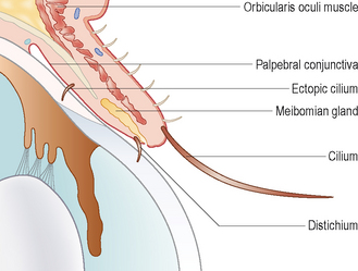
Eyelash abnormalities are very common in dogs but are rarely encountered in cats. Distichia are extra eyelashes along the eyelid margin which originate from slightly abnormal meibomian glands. Ectopic cilia are lashes which grow through the palpebral conjunctiva towards the cornea (Figure 7.1). The typical presentation, if the distichia are clinically relevant, is of wet, itchy eyes. Thus the owner reports the dog’s eyes run constantly and that it rubs them frequently. Some redness is reported but unless corneal ulceration has occurred the eyes are not usually overtly painful. Ectopic cilia more commonly result in corneal ulcers than do distichia. Some breeds such as the Staffordshire bull terrier, English bulldog, boxer, miniature long haired dachshund and cocker spaniel commonly develop distichia, while ectopic cilia are seen in flat coated retrievers and Staffordshire bull terriers more commonly than other breeds. An inherited incidence is clearly present but the exact inheritance has not been evaluated. Typically young dogs are presented from 6 to 18 months.


CLINICAL EXAMINATION


General clinical examination is unremarkable. On ophthalmic examination the aberrant cilia on the eyelid margin can often be seen with the naked eye against the white of the sclera, particularly if they are pigmented. Lashes along the lower lid margin are not immediately so obvious. Increased lacrimation is a feature and Schirmer tear test readings are usually towards the upper end of normal. Some conjunctival hyperaemia is normally present but overt conjunctivitis is not a feature unless ulceration is present. Mild secondary entropion can be present in long-standing cases following chronic blepharospasm and must be differentiated from primary entropion with incidental distichiasis. The application of a drop of topical anaesthesia will alleviate any spastic component to the entropion.


Sometimes the presence of the distichia might be masked by small globules of mucus along the lid margin. Gently bathing the eyes should clear these. Careful examination with good illumination and magnification (+20D on the direct ophthalmoscope is adequate) is required, paying attention to all four eyelid margins. Placing a drop of fluorescein in the eye might highlight the lashes further. Identification of the distichia is not normally difficult – it is deciding whether they are the cause for the dog’s symptoms that is more challenging. Many cases are subclinical, incidental findings, and a thorough examination of the rest of the eyes is required to see if there is another explanation for the lacrimation and irritation. The lashes should be evaluated for position, thickness and number, along with their direction. Even multiple fine lashes might be insignificant if they are not in direct contact with the corneal tear film, whereas one thick, stubby lash directed towards the cornea can be very significant.


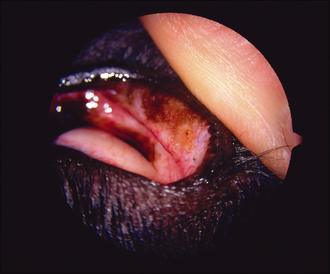
In addition to identifying the lashes, the fluorescein will obviously highlight any corneal ulceration. If this is present, its location should be checked against the position of the distichia. If none are parallel to the ulceration, then they are unlikely to have caused it. Everting the upper eyelid, and checking carefully for any ectopic cilia, is essential if any corneal ulceration is present (Figure 7.2). One or two ectopic cilia can be present, normally close to 12 o’clock on the upper lid, 4–6 mm from the eyelid margin. Their presence is almost always clinically significant. If ulceration is a feature, then a mild reflex uveitis might be noted, but normally there is no intraocular involvement and thus the rest of the ocular examination is unremarkable.


CASE WORK-UP


As mentioned above, the identification of distichia is not particularly challenging, but establishing their relevance can be. As such, a straightforward way to determine their significance is simply to pluck them and see if this alleviates the dog’s symptoms. There should be immediate relief if the lashes are relevant and the owner should report reduced lacrimation and a resolution of the irritation. If this does not occur, the patient requires further detailed ophthalmic evaluation since clearly the distichia were not the cause of the dog’s clinical signs. If the dog is much more comfortable following plucking then when they grow back, usually within 4–6 weeks, one would expect the clinical symptoms to return, further confirming the diagnosis. More permanent treatment options can then be evaluated.


Some dogs will have multiple distichia which cause no problems at all until the dog develops an unrelated painful ocular complaint – perhaps a corneal ulcer as a result of scrapping with another dog or shampoo accidentally splashing in its eye during a bath. The painful eye triggers blepharospasm which changes the angle at which the distichia touch the cornea, causing them to rub and exacerbate the corneal pain. In these instances it is extremely important to decide how relevant the lashes actually are, and plucking them to relieve this secondary discomfort while treating the other eye problem is sensible.


EPIDEMIOLOGY


Distichiasis and ectopic cilia occur on an individual basis in any breed of dog, but some breeds are more commonly affected, such as the Staffordshire bull terrier, English bulldog, cocker spaniel (both English and American), miniature long haired dachshund, boxer and flat coated retriever. The incidence is very high, but the percentage of dogs with ocular disease attributed to the distichia is low. Distichia erupt from meibomian glands. Since these are modified hair follicles anyway the lashes tend to develop from undifferentiated meibomian tissue. Normally the hairs emerge individually but several can come out from one opening (termed districhiasis). They emerge from the meibomian gland orifices as the route of least resistance, but can emerge as ectopic cilia through the palpebral conjunctiva, normally of the upper lid, and this positioning is clinically more severe. The ectopic cilium grows out perpendicular to the cornea and scrapes against it each time the animal blinks, causing a typical linear ulcer in the dorsal cornea.


Sep 10, 2016 | Posted by in SMALL ANIMAL | Comments Off on Eyelash problems – distichiasis and ectopic cilia

Full access? Get Clinical Tree

Get Clinical Tree app for offline access