Chapter 2 Examination of the External Ear Canal
One of the most common ailments of dogs seen in a veterinary practice is ear disease. Approximately 15% to 20% of all canine patients and approximately 6% to 7% of all feline patients have some kind of ear disease, from mild erythema to severe otitis media. Many clients are unaware of their pets’ otitis, and many pets do not show clinical signs of ear disease until it has become quite severe. Determining the cause of ear disease is often a difficult task.
The Normal Ear
Ear Canal
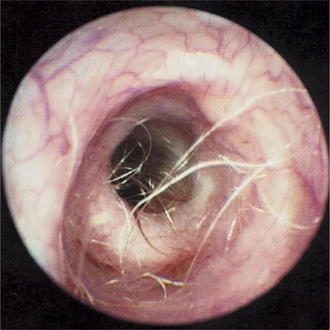
The normal ear canal epithelium should be light pink with small superficial blood vessels visible (Figure 2-1). Small amounts of cerumen coating the epithelium give the surface a glistening appearance. Cerumen is a normal part of a healthy ear and should not be regarded as pathological unless it is excessive. Hairs are seen along the canal, being more numerous in the vertical canal (Figure 2-2).
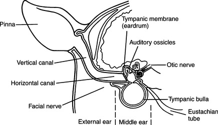
The dog’s ear canal gently bends approximately 75 degrees as it changes from the vertical to the horizontal (Figure 2-3). With the dog in the standing position, the examiner should gently place traction ventrally on the pinna; the ear canal will straighten out, because the normal underlying cartilage is soft and pliable. The otoscope cone should be advanced into the horizontal canal as the canal straightens. This technique enables the horizontal canal to be examined. In the anesthetized patient in lateral recumbency, the examiner can straighten out the canal by lifting the pinna vertically to the point of elevating the entire head.
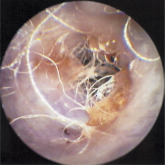
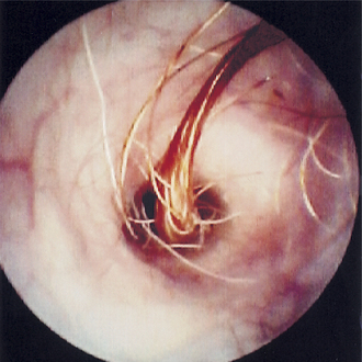
Hairs are almost absent from the horizontal canal in most dogs. However, a clump of long, bristly hairs is often found at the distal end of the horizontal canal and may cause discomfort (Figure 2-4). An accumulation of wax may be seen along the ventral floor of the horizontal canal (Figure 2-5). As the otoscope is advanced in the horizontal canal, the tympanic membrane, if present, should become visible.
Tympanic Membrane
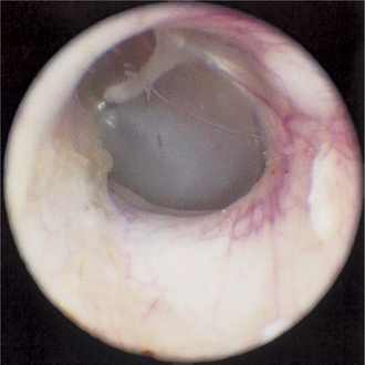
The normal tympanic membrane in both the dog and the cat appears as a thin, transparent to translucent, reflective end of the horizontal ear canal (Figures 2-6 and 2-7). The normal eardrum has been described as having a pale, “rice paper” appearance. The outline of the footplate (manubrium) of the malleus is seen attached to the medial side of the tympanic membrane, frequently bulging outward (pars tensa). The malleus is seen as a thin rectangular white bone originating from the dorsal portion of the tympanic membrane and extending ventrally halfway across the membrane. The malleus is oriented dorsoventrally. The free distal end of the manubrium may have a gentle curve or a “hook” that points rostrally. This feature aids in distinguishing the right ear from the left ear on photographs of the eardrum.

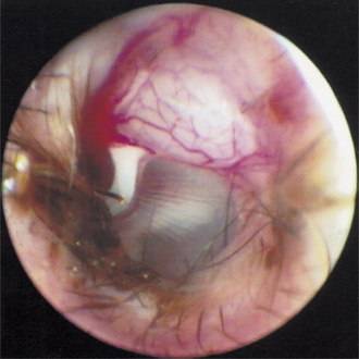
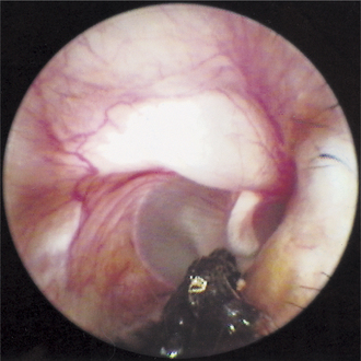
Examination of the dorsal portion of the tympanic membrane (pars flaccida) reveals an opaque, pink or white, loose membrane often containing a network of small blood vessels that extend across the tympanic membrane (Figure 2-8). This “vascular strip” often has an edematous appearance, and this “pretympanic bleb” may obstruct visualization of dorsal portions of the eardrum. In many cases of otitis media, the vascular strip is destroyed, removing from the germinal epithelium of the eardrum the blood supply that is required for the eardrum to heal. If the vascular strip is unaffected by disease or trauma, the damaged eardrum has a much better chance of completely healing with time.