Fig. 12.1.
Representative behavioral testing schedule for MCAO experiment. NS, neuroscore; OF, open field; PPT, paw preference; ORT, object recognition; PA, passive avoidance.
3.3.1 Sensorimotor
Neuroscore: The neuroscore described here for behavioral experiments is somewhat more thorough than other neuroscore methods described in the literature and used in combination with standard histology outcomes (23). The neuroscore covers nine measures of recovery: consciousness, interaction, grooming (eye appearance), respiratory rate, food intake, forelimb strength (ability to grab and hold onto wire cage top), nest building, motor function (standing and walking without leaning or circling), and general activity level. Each of these factors is assigned a different scale ranging from 0 to 3–5, depending on the factor; a maximum score is 23.
The mice are tested for latency to move (see below), weighed, and given subcutaneous fluids as part of the neuroscore testing. Neuroscore is performed on Days 1 and 3 post-operatively, but mice are weighed and given fresh food and fluids daily. Food intake is measured using a dish with a food pellet mixed into mush with water daily.
Outcomes : A control healthy mouse will score 0, while a more impaired animal will score higher. Mice recover well from MCAO and generally by Day 4 or 5 have returned to a normal neuroscore value (<4). Since the animals are being compared to normal via first-hand observation, it is important that the tester has extensive experience including proficient handling skills with both healthy and impaired animals. Our experience is that statistically significant differences in neuroscores across treatment groups are not commonly observed. However, differences are often revealed with more subtle behavioral tasks described here. Nonetheless, performing the neuroscore is important in order to observe gross general health changes and to help habituate the mice to handling by the tester.
Latency to move: The latency to move test measures how quickly an animal moves one body length when placed in an open area. Exposure to the open is stressful to a mouse, and the normal response is to freeze for a few seconds to assess immediate danger and then move to a more protected area (near a wall or crevice). The latency to move test is performed on an open countertop approximately 2 feet square that is designated for this test only. A circle with a radius of 12 cm is drawn in the center of the countertop. The animal is placed by gently sliding it out of the container used to weigh the animals into the center of the circle. Every animal is placed facing the same direction. The animal is timed manually with a stopwatch to measure how long it takes for it to move out of the circle; timing is stopped when all four feet have left the circle. The maximum latency is 60 s. The area is fully cleaned with 70% ethanol after every animal to remove scent trails, which can affect this behavior.
Outcomes : Latency to move in a healthy mouse is roughly 5–10 s. MCAO-injured animals have higher latencies due to apathy or physical inability. Significant improvement (decrease) in latency to move is commonly observed between Days 1 and 3 after MCAO.
Paw preference: Forelimb bias is tested using the paw preference or cylinder test. Focal ischemia generates unilateral damage, resulting in decreased use of the forelimb on the contralateral side to the injury. To track recovery throughout a longer period, this test is performed at Days 3 and 7. Animals are pre-tested on Day 1 to identify any naturally occurring “handedness.” An animal is placed in an upright, clear, plastic cylinder with a diameter approximately equal to the length of the animal’s body. (The restricted space encourages the animal to rear up to explore the cylinder but still gives the animal enough room to turn around freely.) The cylinder sits inside a white plastic box that is similar to the open field test. This blocks out the tester’s movement and provides a familiar environment. Four video cameras are positioned within the box so that all four directions are visualized simultaneously (Fig. 12.2). The video is analyzed in slow motion to ensure accuracy. Only the initial touch is counted each time the mouse rears. A new touch will not be counted until the animal “resets” by returning all four limbs to the floor. If the animal touches the cylinder simultaneously with both forepaws, a count of “both” is entered. The first 20 rears are recorded for each trial.
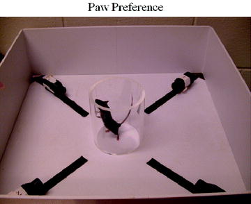
Fig. 12.2.
Paw preference apparatus.
Outcomes : The final score is generally calculated as percent usage of the non-impaired limb. The advantage of this task is that it is sensitive enough to measure impairment at multiple time points for up to 2–3 weeks post-MCAO. However, caution should be taken because over time the mouse may become habituated to the cylinder and stop exploring and rearing. Testing during the dark cycle, when mice are naturally more active, may encourage more rearing behavior. The floor and cylinder are cleaned with 70% ethanol after each trial.
Open field: The open field test is a robust measure of recovery, assessing both anxiety/exploration behavior and recovery of spontaneous locomotor activity. Each animal is tested 1 day before injury and on Day 5 after injury in order to make a direct paired comparison of how the animal has recovered with respect to its prior mobility and willingness to explore. The mouse is placed in a white plastic box measuring 20.5 cm × 20.5 cm × 20.5 cm. An overhead camera records the animal’s movement in four open fields simultaneously. The video is analyzed for average velocity and total distance moved and can also be analyzed for percent time spent near outer walls, as a measure of anxiety. The duration of each trial is 30 min. For all open field trials, each animal is placed in the same box that was used for its original trial to negate any slight differences in lighting or orientation. Each field is cleaned with 70% ethanol after each trial.
Outcomes : All animals should have recovered sufficiently by Day 5 to have equivalent exploration and velocity compared to their baseline scores. If an animal exhibits significantly impaired activity/movement, it will be excluded from all future behavioral testing. The open field task has the added benefit of habituating mice to the field that is also used for novel object recognition.
3.3.2 Cognitive
Novel object recognition: The novel object recognition test is designed to assess hippocampal and cortical damage affecting memory. The test field is a box identical to the one used in the open field test except for two identical objects (e.g., padlock) placed in opposite corners, 1″ away from the walls so that the animals can move fully around them. Objects are chosen in advance to have irregular shapes or defects to appear more interesting and to be approximately the same size and of equal interest to the animals. Objects used frequently are a Masterlock padlock and a plastic clamp, each approximately 1.5″ long. On Day 6 (acquisition/training), the mouse is allowed to explore two identical objects. The trial is stopped when the total exploration time is 38 s, with 10 min maximum duration. Exploration time is measured with two stopwatches (one dedicated to each object), and video is recorded for confirmatory analysis off-line. On Day 7 (test), a novel object (e.g., 1 padlock replaced with plastic clamp) replaces the object the animal spent the least time exploring during acquisition. Time spent exploring the familiar versus novel object is recorded during a 5-min test.
Outcomes : The percent of time spent exploring the new object during the 5-min test trial is used as the final outcome. Healthy animals with intact memory spend approximately 70% of the time exploring the novel object during the test trial. In contrast, injured animals spend significantly less time exploring the novel object, indicative of impaired memory. If an animal does not explore for the required 38 s during the training trial or 16 s during the test trial, then that animal is excluded from the test data.
Passive avoidance: The passive avoidance test is performed on Days 8 and 9 post-injury and is used to assess cognitive function by linking a punishment (short 2-mA shock to the paws) with moving to a preferred environment (dark chamber) during an acquisition/training period. The passive avoidance apparatus (Gemini Avoidance System, SD Instruments) consists of two chambers separated by an automated door. At the start of the acquisition period, both chambers are dark; the animal is placed in the right-hand chamber, and the door is closed. After a 10-s habituation, an overhead light turns on, and the door between the chambers opens. When the animal moves into the dark left-hand chamber, the door closes, and a 2-mA shock is delivered. Ten seconds after the shock is delivered, the animal is removed from the apparatus. Twenty-four hours later, the same protocol is repeated to measure retention of the negative association.
Outcomes : After experiencing a single shock exposure in the dark chamber during the acquisition period, a fully healthy mouse will stay in the non-preferred environment (light chamber) until the maximum latency of 5 min is reached during the test trial 24 h later. Injured animals will enter the dark chamber again on the Day 2, ending the test with their latencies recorded. Animals that do not enter the dark chamber within 40 s on the acquisition day are removed from the test.
3.3.3 Affective
Post-stroke anxiety and depression are common in humans and can be demonstrated in rodents (24, 25). The two most commonly used tasks for assessing anxiety-like behavior after focal cerebral ischemia are the elevated plus maze (EPM) and the open field (OF) test. Both tasks require that the mice are fully mobile and have recovered any gross motor deficits that emerged following stroke.
Elevated plus maze: The EPM apparatus is raised approximately a meter above the floor and consists of two open arms (without walls) and two closed arms arranged in a “+” orientation. The maze is brightly lit to increase contrast between the open and closed arms. All four arms of the maze should be approximately 65 cm long and 5 cm wide. The walls around the closed arms should be approximately 15 cm high. The mouse is placed in the center of the apparatus facing an open arm, and the following measures are recorded: latency to enter arms, duration of time spent in closed and open arms, and frequency of arm entries. The total number of fecal boli excreted during the 5-min test is also recorded.
Outcomes : A significant decrease in the percentage of open arm entries (open arm entries/total arm entries) is indicative of increased anxiety. The rationale is that anxious mice will prefer the relative safety of the dark enclosed arms to the bright open arms. A minimum of six arm entries are necessary for the task to be valid, and significant group differences in the total number of arm entries raise the concern that group differences in general locomotor activity could be a confounding factor in interpreting the EPM data as an index of anxiety-like behavior. An increase in the latency to enter an open arm and an increase in the number of fecal boli expressed can also be indicative of anxiety but tend to be less robust measures than percent open arm entries. Also, increased rearing in the enclosed arms is indicative of increased exploratory behavior.
Open field: In the open field test, anxiety-like behavior is assessed by comparing the amount of activity that occurs at the periphery versus the 100-sq cm zone in the middle of the apparatus. The open field apparatus is 40 cm × 40 cm × 38 cm (l × w × h) and consists of a sound-attenuating chamber with a light and a fan; movement of the mouse within the apparatus is recorded via a digital camera and computer. The bottom of the chamber is covered with clean bedding that is different in texture from the bedding used in the mouse’s home cage. The mouse is placed in the center of the open field chamber at the beginning of the 5-min test. When the test is complete, the bedding is discarded, and all surfaces of the apparatus are cleaned with 70% ethanol.
The three most common tests used to measure depressive-like behavior after focal cerebral ischemia are the sucrose consumption test, the Porsolt swim test, and the tail suspension test.
Outcomes : Decreased percentage of activity in the center of the apparatus (center activity/total activity) indicates increased anxiety-like behavior; the rationale is that anxious rodents avoid such open spaces.
Sucrose consumption test : The sucrose consumption test is based on the observation that rodents find sweet liquids, such as sucrose solutions, rewarding and will drink them preferentially when offered a choice between water and a sucrose solution. A decrease in preference for sucrose is interpreted as anhedonia, a decreased sensitivity to reward. Anhedonia is a core symptom of depression and can be effectively evaluated in rodents.
For this test, the mice must be housed individually. The first step is to familiarize the mice with the water bottles and sucrose solution. The mice should receive all of their water via water bottles beginning 2 weeks prior to stroke. (Be careful to monitor the mice for dehydration if they have been raised on an automatic watering system). One week prior to stroke, remove all water from the cage for the last 6 h of the light cycle. Then, at the onset of the dark cycle, place a bottle containing a pre-measured amount of water and a bottle containing a pre-measured amount of 3% sucrose in the cage. At the end of the first 6 h of the dark cycle, remove the bottles and record the amounts of water and sucrose that were consumed. Repeat this protocol the following night, and calculate the mean water and sucrose consumption for these two nights to determine baseline consumption volumes for each liquid. Following stroke, continue to monitor water consumption. When water consumption has returned to baseline levels, the post-ischemic component of the sucrose consumption test should be initiated using the same protocol as at baseline.


Outcomes : In our experience, mice have a 2:1 preference for sucrose solution over water prior to stroke and no preference for sucrose after stroke. One precaution is to rule out drug effects on taste perception in mice treated with a drug during the sucrose consumption test.< div class='tao-gold-member'>Only gold members can continue reading. Log In or Register to continue
Stay updated, free articles. Join our Telegram channel
Full access? Get Clinical Tree
