Ethylene Glycol

Chapter 78 Ethylene Glycol


METABOLISM


EG is absorbed rapidly from the gastrointestinal (GI) tract (although food slows its absorption time) and is distributed throughout all body tissues. Some of the toxin is eliminated unmetabolized in the urine. Serum concentrations rise rapidly by 1 hour and, in dogs and cats, are typically highest by 3 hours after ingestion. Levels are typically elevated for at least 12 hours but may be undetectable by 48 hours.1


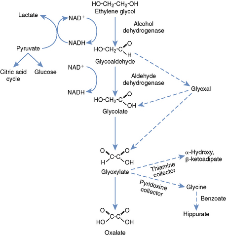
Knowledge of the metabolism of EG is paramount to understanding its toxicity and treatment (Figure 78-1). Metabolism is accomplished primarily in the liver, with a small contribution from the kidneys and stomach. Alcohol dehydrogenase (ADH), an important therapeutic target, converts EG to glycoaldehyde and organic acids. The glycoaldehyde is then converted to glycolate (glycolic acid [GA]), then glyoxylate (also known as glyoxylic acid). Because the metabolism of GA is rate limiting, this metabolite achieves much higher concentrations in the blood than any of the others. Glyoxylate is converted primarily to oxalate, but additional end products include glycine, formic acid, hippuric acid, oxalomalic acid, and benzoic acid. The oxalate formed from glyoxylate combines with calcium to form the characteristic calcium oxalate crystals that are deposited throughout the body, but primarily in the kidneys.1,3


TOXICITY AND SYSTEMIC EFFECTS


Compared with its metabolites, EG is relatively nontoxic, but it is a potent central nervous system (CNS) depressant and GI irritant. Its metabolites are highly toxic and have a myriad of deleterious systemic sequelae. Glyoxylate and glycoaldehyde are considered the most toxic of the metabolites on a per weight basis. However, because of its longer half-life and greater systemic accumulation, GA is thought to be the major mediator of in vivo toxicity.1


CNS depression can occur within 30 minutes of EG ingestion and typically manifests as depression, incoordination, and ataxia. With more severe intoxications, paresis, somnolence, seizures, and coma can be seen. It is extremely important to associate these clinical signs with EG toxicity because these are some of the earliest manifestations of intoxication, and thus recognition can lead to early and effective treatment. The CNS signs may abate 12 hours post ingestion in dogs, and patients often appear to have “recovered” for a brief time. The CNS effects are thought to result from a combination of direct effects from aldehyde metabolites, hyperosmolality, and metabolic acidosis. Glycoaldehyde adversely affects respiration, glucose, CNS amine concentrations, and serotonin metabolism. For this reason, CNS toxicity correlates better with GA than EG concentrations.1,5


GI manifestations are highly nonspecific. Vomiting can occur immediately post ingestion from direct gastric mucosal irritation. It can also be seen as serum osmolality rises, thus stimulating the chemoreceptor trigger zone. Deposits of calcium oxalate and focal bleeding have been found in the stomach at necropsy. Severe GI disturbance is often a common late sequela, as acute renal failure (ARF) progresses to uremia.1


Cardiorespiratory effects are a much less prominent manifestation of EG poisoning in dogs and cats compared with humans. Tachycardia and tachypnea are seen more commonly in dogs than in cats. Hypocalcemia and metabolic acidosis can both lead to arrhythmogenesis and decreased inotropy. Myocardial calcium oxalate deposition is occasionally documented during necropsy.1,3


ARF is the most commonly associated and best documented systemic manifestation of EG intoxication in small animal medicine. ARF typically occurs 24 to 72 hours after ingestion in dogs but can occur as early as 12 hours after ingestion in cats. Unlike with some other nephrotoxicants such as nonsteroidal antiinflammatory drugs and aminoglycosides, EG is often associated with oliguric or anuric ARF. Mechanisms of nephrotoxicity are still incompletely understood. Calcium oxalate crystal deposition within the tubules and proximal tubular epithelium plays a role, but in humans the degree of renal damage is not correlated with the degree of crystal formation.


Direct renal tubular epithelial cytotoxicity from EG metabolites likely plays a significant role in the acute tubular necrosis (ATN). Glyoxylate and glycoaldehyde are highly nephrotoxic in vitro; however, GA is thought to be one of the main mediators of acute tubular necrosis that is seen in clinical cases. In humans, GA concentrations correlate with progression to ARF, and levels greater than 10 mmol/L are highly predictive of ARF. Duration of exposure to cytotoxic metabolites is thought to influence the degree of renal injury as well. Polyuria and polydipsia (PU/PD) can be marked in dogs, often develop shortly after EG ingestion, and are secondary to an osmotic diuresis.1,3,5


DIAGNOSIS


As will be discussed in the next section, early treatment of EG intoxication is extremely important to achieve a successful clinical outcome. Early diagnosis is therefore key. Clinical signs are dose dependent and related to those caused by unmetabolized EG, followed by those caused by the toxic metabolites. Initial clinical signs are often apparent 30 minutes after EG ingestion and include nausea and vomiting, CNS depression, ataxia, decreased proprioception, lower motor neuron signs to the limbs, muscle fasciculations, hypothermia, and polyuria and polydipsia. The CNS changes typically abate after 12 hours in dogs, but cats often remain severely depressed. Clinical signs of ARF (24 to 72 hours in dogs and 12 to 24 hours in cats) may include coma and depression, anorexia, vomiting, seizures, ptyalism, and oral ulcerations. Anuria is commonly seen 72 to 96 hours after ingestion, and the kidneys are often painful and swollen upon palpation in cats.


The first clinicopathologic finding seen in patients with EG ingestion is a rise in the osmolal gap (OG) (Box 78-1). This can occur as early as 1 hour after ingestion in cats and dogs and typically peaks by 6 hours in dogs. The OG remains elevated for approximately 18 hours after ingestion of EG. The OG correlates well with EG concentration in both humans and dogs, and in human intoxications the deviation from a particular laboratory’s “normal” OG (typically around 10 mOsm/kg H2O; up to 150 mOsm/kg with EG toxicity) can be multiplied by 6.2 to estimate EG concentration in mg/dl. Data from dogs again show a high correlation between OG and EG concentrations, but this formula appears to be less reliable in this species. Serum osmolality should be measured by freezing point depression to accurately calculate the OG.1,3-6


Stay updated, free articles. Join our Telegram channel

Sep 10, 2016 | Posted by in SMALL ANIMAL | Comments Off on Ethylene Glycol

Full access? Get Clinical Tree

Get Clinical Tree app for offline access