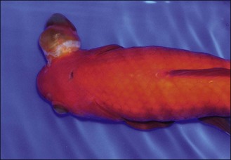
26 Enucleation of an eye in a goldfish
Case History
CLINICAL TIPS
Transport of fish to veterinary clinic
• A container large enough to permit some swimming is necessary. Usually this is a leak-proof plastic box or bucket, but may need to be a thick plastic bag within a firm container such as a polystyrene box.
• To avoid undue stress to the animal with water changes, the fish should be transported in water from its enclosure.
Clinical Examination
The patient was initially assessed in the presenting container:
CLINICAL TIPS
Clinical examination of fish
• Observation of the fish in water provides useful information, including assessment of the animal’s mobility and respiratory movements.
• In some cases it is necessary to perform a brief conscious examination with the fish out of the water:
• Some of the examination may be performed with the fish in water, or it may be raised into the air for a brief period.
• Handling is stressful for fish, and may result in physical damage. Although this may appear minor, for example with the loss of a few scales, this may provide an entry portal for infection that may progress to septicaemia and death. Great care should therefore be taken when handling specimens. Nets should be free of holes that may catch a fin, and of abrasive surfaces.
Case Work-Up
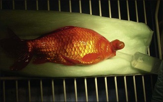
The goldfish was anaesthetized using tricaine methane-sulfonate (MS-222). Once induced, the fish was positioned in lateral recumbency on a wet incontinence pad. Water was dripped over the fish’s body to keep the integument moist. Anaesthesia was maintained by slowly syringing a 100 mg/ml solution of buffered MS-222 into the oral cavity using a catheter-tip syringe (Fig. 26.2).
A full clinical examination was performed:
• Right eye was enlarged. The cornea was opaque so intra-ophthalmic examination was not possible. No discharge was present
CLINICAL TIPS


