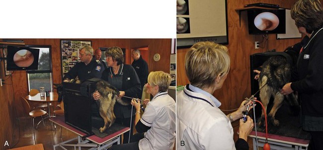
Endoscopic Transcervical Insemination

Chapter 206


Endoscopic Transcervical Insemination


In many species frozen-thawed semen must be deposited into the uterus rather than the vagina to achieve good pregnancy rates and litter size. The options available to achieve intrauterine semen deposition are surgical or transcervical insemination (TCI). Many veterinarians choose to deposit semen surgically into the uterus of the bitch because the surgery is easy to perform. However, surgery is an invasive insemination technique and has risks associated with general anesthesia and surgery, which restrict the veterinarian to a single insemination. In a number of European countries the surgical insemination procedure is not permitted because it is considered ethically unacceptable. Furthermore, many owners and veterinarians prefer a noninvasive, nonsurgical option.


Dr. Marion Wilson developed the noninvasive TCI technique using a rigid endoscope specifically to deposit frozen-thawed semen into the uterus of the bitch (Wilson, 2001). Research of this technique resulted in identification of several features relating to the anatomy of the canine reproductive tract. These points are relevant to endoscopic TCI and are considered first, followed by a description of relevant procedures for TCI.


Anatomy


The cranial vagina (paracervix) is dominated by the dorsal median fold (DMF), which significantly reduces the vaginal lumen in the approach to the cervix. The restricted vaginal lumen limits the diameter of equipment that can be passed through the area. This feature, together with a particularly long vagina in the bitch, limits the number of endoscopes suitable for the technique. The DMF ends cranially at the vaginal portion of the cervix, which exists as a distinct tubercle (cervical tubercle). The paracervix is limited cranially by the fornix, which is a slitlike space cranioventral to the cervical tubercle. It appears as a blind end when viewed through the endoscope and is another important landmark. The cervix lies diagonally across the uterovaginal junction with the canal of the cervix directed craniodorsally from the vagina to the uterus. Consequently the external os is located ventrally in the cervical tubercle.


Equipment


The endoscope initially identified as meeting the criteria with regard to length and diameter was a rigid, extended length cystourethroscope (Table 206-1). An 8 Fr urinary catheter is used for insemination in the majority of bitches using this endoscope, although a 6 Fr gauge sometimes is required in small or maiden bitches. More recently, a longer (43 cm) and thinner (graduated 9.5 Fr to 13.5 Fr) rigid renourethroscope has been developed for the TCI procedure in bitches. A specially designed 70 cm × 5 Fr catheter (Minitube) with removable stylet is used for insemination of most bitches. Occasionally a 4 Fr catheter is required in small or toy breeds that can have a smaller os and cervical canal.


The longer, thinner endoscope has a number of advantages. First, the significantly narrower shaft greatly facilitates passage under the DMF, which is often a tight and narrow space. This feature is particularly beneficial in maiden bitches and certain individual bitches that can have a narrow vaginal canal. The longer endoscope has eliminated any limitations previously associated with insemination of large and giant breed bitches. However, despite the increased length of the new endoscope, it still can be used with relative ease in toy breeds. Finally, the renourethroscope is technically easier to use in regard to manipulation and catheterization of the os cervix compared with the cystourethroscope.


Furthermore, a “TCI Shunt System” (Minitube) has recently been developed to be used in combination with the renourethroscope for intrauterine insemination of bitches. This device consists of a shunt made of metal and a Foley catheter 55 mm or 105 mm in length. The shunt has multiple functions. Firstly the Foley component creates a tight seal in the vagina, thus eliminating air loss from the vaginal space during air insufflation. This allows the TCI endoscope to pass easily through the caudal vagina and under the dorsal median fold to the cervix. The shunt enhances stabilization of the TCI endoscope and fixation and catheterization of the cervical os. The Foley component also causes stretching of the vaginal wall, stimulating release of local oxytocin and resultant vaginal and uterine contractions, which facilitate sperm transport to the site of fertilization.


A specially designed platform is used to restrain the bitch in the standing position. The platform provides a tie point to the dog’s collar and an abdominal support to restrict sideways movement and discourage any attempt to sit (Figure 206-1). A hydraulic chair together with a hydraulic table for the platform ensures the optimum position of the bitch relative to the operator during the procedure. This is important particularly when the endoscope is used without a camera. Bitches in estrus exhibiting standing behavior show excellent tolerance to the technique and sedation is rarely necessary.


Technique


The endoscope is introduced through the vulva and into the vestibule, taking care to avoid the clitoris and urethral orifice. It is advanced through the vaginal folds by observing the direction of the vaginal lumen. Air insufflation allows greater visualization of the vaginal lumen so that it can be “followed” easily to the cervix. Air insufflation is especially necessary for the renourethroscope because it does not have a sheath. In proestrus and early estrus, the rounded vaginal folds can make advancing the endoscope towards the cervix more difficult because the folds tend to fill the lumen. As estrus progresses, dehydration and crenulation of the folds result in a less obstructive lumen. The DMF and crescentlike lumen of the cranial vagina represent important landmarks in locating the cervical os. At this point, the vaginal lumen can be narrow in some bitches, requiring manipulation of the endoscope to the widest space. This may result in the endoscope being pushed to one side of the DMF rather than continuing ventrally under the DMF. The ventral location of the cervical os means that it is not immediately obvious and the scope has to be directed under the cervical tubercle until the os, located in the center of a rosette of furrows, can be identified. The catheter is advanced into the cervical os by manipulation of the endoscope and catheter. The rigidity of the endoscope is used to move the cervical tubercle, line up the os, and change the angle of the canal.


Once the tip of the catheter is introduced into the cervical os, it is advanced steadily using a clockwise twisting motion to aid its passage through the cervical canal. The catheter should be passed through the cervix as far as it will go without force. Use of 5-cm marked graduations on the catheter is helpful in determining how far the catheter has advanced. The semen is inseminated slowly under continual observation to ensure that no significant backflow of semen into the vagina occurs. If this happens the catheter should be relocated, either further in or back slightly, and insemination started again.

< div class='tao-gold-member'>

Stay updated, free articles. Join our Telegram channel

Jul 18, 2016 | Posted by in PHARMACOLOGY, TOXICOLOGY & THERAPEUTICS | Comments Off on Endoscopic Transcervical Insemination

Full access? Get Clinical Tree

Get Clinical Tree app for offline access