Electrodiagnostics for Retinal Disease

CHAPTER 146 Electrodiagnostic Testing for Retinal Disease


Evaluation of the ocular fundus is an important part of ophthalmic examination in horses. However, retinal disease might not always manifest with visible fundic lesions, and it may be difficult to determine objectively the impact of a retinal lesion on visual performance. Several functional tests are available to evaluate or estimate a horse’s visual performance, including assessment of the menace response and use of obstacle courses. Other methods help to evaluate portions of the visual pathways, but they do not assess vision per se (i.e., conscious visual perception) because a positive response does not depend on relaying of the light stimulus through the visual cortex; these tests include assessment of the pupillary light reflex (PLR) and dazzle reflex. Although retinal function is required for a positive test outcome, none of these evaluation methods can specifically measure retinal function because several parts of the visual system are involved. The electroretinogram (ERG) is the only method that specifically and objectively measures retinal function; it is the recorded total electrical response of the retina to a light stimulus. The purpose of this chapter is to introduce practitioners to the use of ERG in horses and discuss the indications for ERG recording.


TECHNICAL ASPECTS OF ELECTRORETINOGRAPHY


The principles of ERG recording are similar to those of other clinical electrophysiologic techniques, such as electrocardiography. In contrast to electrocardiography, ERG requires not only a recording system, but also a stimulating system. The retina is stimulated with brief light flashes, and the retinal response to these light stimuli is recorded from the ocular surface as the ERG. The components of the ERG have been well characterized; wave amplitudes and implicit times or latencies can be measured, permitting objective assessment of retinal function.


Preparation of the Horse


The first step in preparation of the horse for ERG recordings consists of dilation of the pupils with a short-acting mydriatic such as 1% tropicamide solution. Mydriasis is important for avoiding uncontrolled variation in the amount of light reaching the retina. If the pupil is subject to constriction during testing, results acquired at different sessions or from different horses cannot be compared.


Although ERGs are recorded in awake humans, most animals must be immobilized to some degree to avoid excessive head movement once the light stimulus and the recording electrodes are in place. In small animals, sedation is used for short recording protocols that are used to detect or rule out advanced loss of retinal function. Diagnosis of early or mild retinal degeneration necessitates longer recording protocols with the animal under general anesthesia. Because general anesthesia is generally not justified in horses for the detection of abnormal retinal function, protocols have been developed for ERG in standing horses under sedation. Intravenous injection of detomidine hydrochloride is very useful for obtaining ERG recordings. For longer protocols the duration of sedation can be extended by administration of a constant-rate infusion of detomidine. It is advantageous to restrain the horse in stocks with a padded headrest during recording.


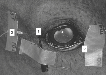
Before an electrode can be placed on the cornea, topical anesthesia of the ocular surface with proparacaine or tetracaine is usually required. Placement of an auriculopalpebral nerve block is recommended to immobilize eyelid musculature during recording. Depending on the type of corneal electrode used, it may be necessary to open the palpebral fissure either manually or by use of a lid speculum so that the amount of light entering the eye is not decreased by the eyelids.


For analysis of rod and cone photoreceptor-mediated retinal function, a complete protocol should include ERG recordings obtained under dark or scotopic and bright or photopic light conditions. At least 20 minutes should be permitted for dark adaptation, and 5 to 10 minutes should be permitted for light adaptation. Because of the long adaptation times and special equipment required, ERG for horses is offered only at selected specialty clinics and is performed mostly by veterinary ophthalmologists.


May 28, 2016 | Posted by in EQUINE MEDICINE | Comments Off on Electrodiagnostics for Retinal Disease

Full access? Get Clinical Tree

Get Clinical Tree app for offline access