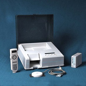
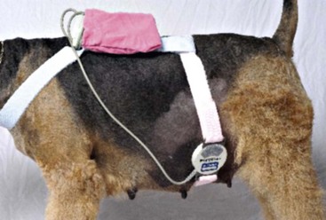
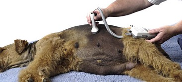
Dystocia Management

Chapter 208


Dystocia Management


Although many dogs and cats deliver in the home or kennel/cattery setting without difficulty, requests for veterinary participation in the field of veterinary obstetrics have become more common. The increased financial and emotional value of stud dogs and toms, brood bitches and queens, and their offspring to the dog and cat fancier makes the preventable loss of even one neonate undesirable. Breeding colonies in academic, scientific, and industrial facilities must maximize neonatal survival for financial and ethical reasons.


Veterinary involvement in obstetrics has several goals: to increase neonatal viability (minimizing offspring born dead from the difficulties in the birth process), to minimize morbidity and mortality in the dam, and to contribute to better survival rates of neonates during the first week of life. Neonatal survival is related directly to the quality of labor. Optimal management of parturition requires an understanding of normal labor and delivery in the bitch and queen, as well as the clinical ability to detect and treat abnormalities in the delivery process.


Dystocia is defined as difficulty in the normal vaginal delivery of a neonate from the uterus. Dystocia must be detected in a timely fashion for medical or surgical intervention to improve outcome. In addition, the cause of dystocia must be identified for the most appropriate therapeutic decisions to be made.


Normal Gestation in the Bitch


Clinicians commonly are asked to ascertain if a bitch is at term pregnancy and ready chronologically to deliver a litter and even to intervene if labor has not begun when anticipated. Because there are no well-documented, predictable, and safe labor induction protocols for the bitch, this generally means a cesarean section.


Prolonged gestation is a form of dystocia. Normal gestation in the bitch is 56 to 58 days from the first day of diestrus (detected by serial vaginal cytologies, defined as the first day that cytology returns to 50% or fewer cornified/superficial cells) or 64 to 66 days from the initial rise in progesterone from baseline (generally between 1.0 and 2.5 ng/ml), which equates to the day of the luteinizing hormone (LH) surge, or 58 to 72 days from the first instance that the bitch permitted breeding. Predicting gestational length without any prior ovulation timing is difficult because of the disparity between estrual behavior and the actual time of conception in the bitch and the length of time semen can remain viable in the bitch reproductive tract, often up to more than 7 days. As a result, breeding and conception dates do not correlate closely enough to permit highly accurate prediction of whelping dates. In addition, clinical signs of term pregnancy are not specific: the radiographic appearance of fetal skeletal mineralization varies at term; fetal size varies with breed and litter size, making ultrasonographic gestational aging imprecise; and the characteristic drop in body temperature preceding stage I labor (typically less than 100° F) varies and may not be detected in all bitches.


Breed, parity, and litter size also can influence gestational length. Astute dog owners and clinicians may be able to detect subtle signs of impending delivery, such as relaxation of the perineum, mammary engorgement, or a change in the appearance of the gravid abdomen, but these are not sensitive or specific. The inability to manage clinically prematurity in the canine (primarily because of lack of surfactant) effectively makes excessively early delivery undesirable. Unfortunately an overly conservative (delayed) approach resulting in intrauterine fetal death also is undesirable.


Bitches typically enter stage I labor within 24 hours of a decline in serum progesterone to below 2.0 ng/ml, which occurs in conjunction with elevated circulating prostaglandins and is associated commonly with a transient drop in body temperature usually to less than 100° F. Monitoring serial progesterone levels for gestational length is problematic because in-house kits enabling rapid results are inherently less accurate between 2 and 5 ng/ml. Commercial laboratories offering quantitative progesterone by chemiluminescence enzyme immunoassay typically have a 12- to 24-hour turnaround time, which is not rapid enough to enable decisions about an immediate indication for obstetric intervention. Progesterone levels can drop precipitously over an 8- to 12-hour period; thus results are helpful in supporting the decision to perform an elective cesarean section only if the level drops below 2.0 ng/ml. Some bitches experience a typical drop in progesterone level but still do not have normal labor. Clearly it is beneficial to obtain information about ovulation timing, minimally by determining the onset of cytologic diestrus, for accurately evaluating gestational length at term. The clinician should avoid electively delivering puppies younger than 62 gestational days (days from the LH surge), unless an indication of impending fetal demise or maternal morbidity is present. Doppler or ultrasonographic fetal heart rate monitoring may be helpful; if fetal heart rates are consistently less than 150 beats/min, fetal stress is likely.


Normal Labor and Delivery


Stage I labor in the bitch and queen normally lasts from 12 to 24 hours, during which time the uterus has myometrial contractions of increasing frequency and strength associated with cervical dilation. No abdominal effort (visible tenesmus) is evident during stage I labor. Bitches and queens may exhibit changes in disposition and behavior, becoming reclusive and restless and nesting intermittently, often refusing to eat, and sometimes vomiting. Panting and trembling may occur. Vaginal discharge typically is clear and watery.


Normal stage II labor is defined to begin when external abdominal efforts can be seen accompanying myometrial contractions to culminate in the delivery of a neonate. Presentation of the fetus at the cervix triggers the Ferguson reflex, promoting the release of endogenous oxytocin from the hypothalamus. Typically these efforts should not last longer than 1 to 2 hours between fetuses, although this varies greatly. The entire delivery can take between 1 and more than 24 hours; however, normal labor outcome is associated with shorter total delivery time and intervals between neonates. Vaginal discharge can be clear, serous to hemorrhagic, or reddish brown in queens or green (uteroverdin) in bitches. Typically dams continue to nest between deliveries and may nurse and groom neonates intermittently. Anorexia, panting, and trembling are common.


Stage III labor is defined as the delivery of the placenta. Dams typically vacillate between labor stages II and III until the delivery is complete. During normal labor all fetuses and placentae are delivered vaginally, although they may not be delivered together in every instance. Occasionally placentae are delivered hours after delivery of all of the offspring is complete. Dams typically consume placentae, making client counts potentially inaccurate.


Detecting Abnormalities in Canine and Feline Labor


The standard approach to labor management in companion animals has involved subjective monitoring of the dam’s behavior, rectal temperature (in bitches), progression of labor, and the physical condition of the neonates. Little accurate, timely, and evidence-based information is made available to the clinician concerning actual uterine activity or prepartum fetal viability with this methodology. Telephone consultations between the veterinarian and breeder or kennel manager usually entail interpretation of indirect information such as time between deliveries, color of vaginal discharge, presence and nature of externally visible tenesmus, and occurrence of stillborn offspring. Although generally acceptable (associated with a favorable outcome) for the uneventful delivery in a young, healthy dam, parturition associated with fetal and maternal morbidity and mortality is familiar to most clinicians in reproductive practice. For these cases, improved diagnostic tools are desirable, such as those used in human obstetrics. Clinical information about the onset, duration, and progression of the stages of labor, frequency and strength of myometrial contractions, progression of delivery of fetuses into the uterine body and vagina for birth, and physical status of the fetuses is helpful for obstetric decision making. Evaluating the metabolic status of dams presented for dystocia is important if hypocalcemia or hypoglycemia is suspected but neither commonly is detected.


Recently a system for monitoring labor and fetal viability in the bitch or queen has become available commercially (Veterinary Perinatal Services, Inc.). This system is intended for use by veterinarians in the clinical setting when evaluating a bitch or queen in labor or by dog or cat fanciers or kennel or cattery managers with veterinary supervision in the home or kennel/cattery setting. The Veterinary Perinatal Service (VPS) design is based on labor monitoring systems used routinely in human obstetrics. The uterine monitoring system consists of a tocodynamometer (sensor), a recorder, and a modem (Figure 208-1). The uterine sensor detects changes in intrauterine and intraamniotic pressures. The sensor is strapped over a lightly clipped area of the dam’s caudolateral abdomen using an elasticized strap. The sensor’s recorder is worn in a small backpack placed over the caudal shoulder area in medium and large bitches or adjacent to this area in smaller bitches or queens (Figure 208-2). Dams should be at rest in the nesting box, cage, or a crate during the monitoring sessions. The monitoring equipment is well tolerated. Subsequent to each recording session, data are transferred from the recorder to the service center using a modem with the telephone.


Fetal heart rate assessments can be performed easily with either real-time ultrasound or a handheld Doppler unit. Normal fetal heart rates generally range above 220 beats/min. Normal canine and feline fetuses at term have heart rates at least twice the maternal rate. Fetal distress is evident by persistent fetal bradycardia of 120 to 150 beats/min. In a model of impaired oxygen delivery, heart rates in fetal dogs were recorded during hypoxic episodes; they showed that decelerations were an early sign of fetal hypoxia.


Fetal heart rate monitoring, performed with either a real-time ultrasound scanhead (5 to 7.5 MHz) or a handheld Doppler unit, is best used with dams in lateral recumbency, using acoustic coupling gel (Figure 208-3). Visualization of the fetal cardiac valve motion enables estimation of the heart rate with real-time ultrasound. Directing the handheld Doppler perpendicularly over a fetus results in a characteristic amplification of the fetal heart sounds, distinct from maternal arterial, digestive, or cardiac sounds, enabling audible determination of fetal heart rates. The presence of fetal distress is reflected by sustained deceleration of the heart rates. Decelerations associated with uterine contractions suggest mismatch between the size of the fetus and the birth canal or fetal malposition or malposture. Malpresentation is generally not a problem in canine or feline deliveries because approximately 50% are delivered in caudal longitudinal presentation. Transient accelerations occur with normal fetal movement.


< div class='tao-gold-member'>

Stay updated, free articles. Join our Telegram channel

Jul 18, 2016 | Posted by in PHARMACOLOGY, TOXICOLOGY & THERAPEUTICS | Comments Off on Dystocia Management

Full access? Get Clinical Tree

Get Clinical Tree app for offline access