Fig. 26.1
Scanning electron micrographs of freeze-fractured renal corpuscles of living mice under normal blood flow condition, as prepared by IVCT. (a) Interdigitating foot process es are covering the outer capillary loop surface. P podocytes, F foot processes, U urinary space, B Bowman’s capsule. Bar: 5 μm. (b) At higher magnification, surface contours of foot processes (F) with various slit spaces are arranged almost in parallel with each other. Bar: 1 μm. (c) Filtration slits (arrows) are also seen to be open between foot processes (F) under the normal blood flow condition. BM basement membrane. Bar: 0.5 μm
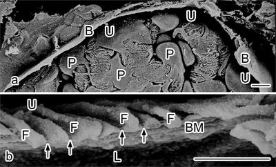
Fig. 26.2
Scanning electron micrographs of freeze-fractured renal corpuscles of mice under the heart-arrest condition , as prepared by IVCT. (a) The urinary space (U) is almost collapsed, which is also covered by outer Bowman’s capsule (B). Bar: 5 μm. (b) A freeze-fractured capillary loop is seen, and foot process es (F) with narrow filtration slit s (arrows) are attached to the basement membrane (BM). Bars: 1 μm. L capillary lumen, U urinary space
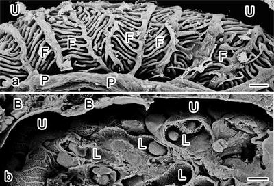
Fig. 26.3
Scanning electron micrographs of freeze-fractured renal corpuscles of living mice under the aorta ligation condition, as prepared by IVCT. (a) Interdigitating foot process es (F) are extended to become slender, covering the outer surface of blood capillary loops. P podocyte, U urinary space. Bars: 1 μm. (b) The lumens of freeze-fractured capillary loops (L) are also more widely open with dilatation of the urinary space (U). B Bowman’s capsule. Bar: 5 μm
26.4 Dynamically Changing of Foot Processes for Filtration Functions
It has been known that glomerular capillary loop s of living mouse kidney s are perpetually stretched by hydraulic pressures in blood capillaries , whereas those in excised kidneys receive no such stretching forces. Filtration slits between foot process es of podocytes were reported to be widely open to facilitate passage of the glomerular filtrate in vivo [3]. It has also been reported that the foot processes are rich in contractile proteins, such as actin, myosin, and α-actinin, and capable of such actin-mediated movement [11]. Through this active movement, the podocytes may regulate the glomerular filtration rate , thereby influencing hydraulic pressures across the glomerular basement membrane , as reported before [3]. Therefore, the blood pressures must be steadily maintained at the time of exposure to quick freezing with IVCT, and the dynamically changing filtration slit s are found to be wider under normal blood circulation than the heart-arrestcondition . The glomerular hydraulic pressure s might be partially regulated by the relative width of the filtration slits. Therefore, the total width of the filtration slits plays an important role in providing a porous theory that controls hydraulic conductivity and water flow [3, 12].
Stay updated, free articles. Join our Telegram channel

Full access? Get Clinical Tree


