Diuretics

Chapter 180 Diuretics


INTRODUCTION


Disturbances in the regulation and balance of fluid and electrolytes are very common, and they contribute significantly to the morbidity and mortality of animals treated in the critical care setting. Fluid and solute excesses are often corrected with diuretics, a heterogeneous group of drugs acting on various segments of the nephron, where they block the reabsorption of water and solutes and promote their urinary excretion. The correct assessment of electrolyte and mineral disorders is hampered by their compartmentalization, and correction of their serum concentrations is sometimes better achieved by translocation into the proper compartment.


The appropriate use of diuretics in the critical care patient requires a careful clinical and laboratory assessment and a good understanding of the underlying disease and pathophysiology in order to define clear therapeutic goals for the various fluid compartments, electrolytes, and minerals, and to choose the most appropriate diuretic, its route of administration, and dosage. Because of the complex disease processes of most critically ill animals, the limitations of their clinical assessment, and the limited data from clinical studies in small animals, this therapy remains often empiric and based on pathophysiologic justifications and clinical experience rather than on objective experimental data. The therapeutic monitoring should therefore aim to more objectively assess treatment success and anticipate or recognize side effects.


PHYSIOLOGY OF DIURESIS AND ANTIDIURESIS


One of the main characteristics of the kidney function is its ability to regulate the excretion of water and most individual solutes independently of each other.1 In the normal animal the rate of urine excretion (diuresis) depends mostly on renal handling of water and thus on the concentration of antidiuretic hormone (ADH, vasopressin). ADH production is increased in response to elevated plasma osmolality, hypovolemia/hypotension and, to a lesser extent, as a response to nausea and increased angiotensin II concentration. ADH production is suppressed and diuresis is increased by atrial natriuretic hormone and ethanol.1


To exert its antidiuretic function, ADH requires a functional tubular system, a medullary concentration gradient of sodium and urea, and a functional ADH-receptor system to use this gradient. Failure of these mechanisms results in an inappropriately increased diuresis. Two additional diuretic mechanisms are involved in pathologic conditions: (1) pressure natriuresis, a negative feedback involved in hypervolemic hypertensive states leading to increased natriuresis and restoration of normovolemia and normotension, and (2) osmotic diuresis, a passive diuretic mechanism resulting from abnormal urinary concentrations of osmotically active solutes such as glucose or sodium.1


Therefore increased diuresis can be achieved therapeutically through exogenous loading with water or salt, administration of poorly reabsorbed solutes, and pharmacologic inhibition of the tubular reabsorption mechanisms of sodium or water. Depending on the mechanisms involved, the diuretic effect will be associated with extracellular fluid (ECF) volume expansion (hypervolemic diuresis) or depletion (hypovolemic diuresis).


PHARMACOLOGY


Diuresis can be induced osmotically or, more commonly, by pharmacologic blockade of sodium reabsorption at various sites along the nephron. The basic rule is that, although they can modulate a greater bulk of sodium, efficacy of proximal diuretics may be overcome by distal compensatory increases in sodium reabsorption in the loop of Henle. The efficacy of distal diuretics on the other side is limited by the small portion of sodium actually reaching the distal tubule. Diuretics acting at the loop of Henle are thus most effective because of the large amount of filtrate delivered to this site and the lack of an efficient reabsorptive region beyond their site of action.2


Diuretics are grouped according to their mechanism of action and they include, in order of their renal tubular target, osmotic diuretics, carbonic anhydrase (CA) inhibitors, loop diuretics, thiazide diuretics, aldosterone antagonists, and other potassium-sparing distal diuretics (Table 180-1). Mannitol and furosemide are used most frequently in the critical care setting; consequently the other diuretics will be mentioned here only briefly. Usual dosage recommendations are summarized in Table 180-2. In using diuretics it is important to note that fluid therapy should be adjusted closely to the desired goals of the global therapy. For example, if the therapeutic goal is a depletion of the ECF with furosemide in an animal with CHF, it does not make sense to administer intravenous fluids concomitantly. Partial free water replacement with 5% dextrose in water may be an exception in this scenario.


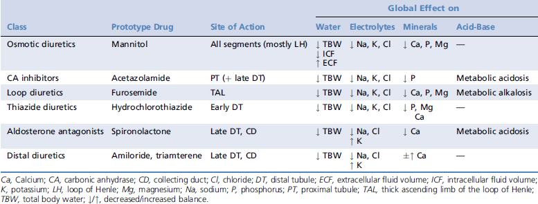
Table 180-2 Dosages of Most Commonly Used Diuretics


Drug Indication Dosage
Mannitol Renal failure 0.25 to 1 g/kg IV q4-6h
CRI 1 to 2 mg/kg/min when diuresis instituted
  Glaucoma 1 to 3 g/kg IV once
  Cerebral edema 1 to 1.5 g/kg IV once
Acetazolamide Glaucoma 50 mg IV once, then 2 to 10 mg/kg q8-12h PO
7 mg/kg PO q8h in the cat
Furosemide Diuretic 0.5 to 4 (max 8) mg/kg IV, IM, SC, PO q8-12h
CRI 2 to 15 μg/kg/min
Hydrochlorothiazide Diuretic 0.5 to 5 mg/kg PO q12-24h
Spironolactone K-sparing diuretic 1 to 4 mg/kg PO q12-24h
Amiloride K-sparing diuretic 0.1 to 0.3 mg/kg PO q24h
Triamterene K-sparing diuretic 1 to 2 mg/kg PO q12h

CRI, Constant rate infusion; IM, intramuscular; IV, intravenous; K, potassium; PO, per os; SC, subcutaneous.


All diuretics except spironolactone reach their tubular sites of action through the urinary space. Mannitol is freely filtered in the glomeruli, and the other highly protein-bound diuretics are secreted actively through the organic acid and organic base pathways into the proximal tubule.2 This explains the decreased efficacy of most diuretics in animals with renal disease. However, the impaired tubular secretion and delivery to the site of action can be compensated partially by a progressive titration to higher plasma concentrations. In animals with proteinuria and nephrotic syndrome, the serum diuretic concentration remains low as a result of hypoproteinemia and results in decreased tubular secretion of the diuretic that is then partially neutralized by binding to urinary proteins. Dose and frequency adjustments can partially compensate for this and provide sufficient concentrations of the active drugs at the site of action.2,3 Serial measurements of urinary electrolytes can provide a more objective assessment of diuretic efficacy to help guide therapeutic decision making, such as dosage adjustments and combining diuretics from different classes.


Tolerance and inefficacy of diuretic therapy can happen after a single dose as a result of depletion of the ECF. In the long term, hypertrophy of the distal nephron reflects an increased compensatory solute reabsorption of the distal sites to compensate for proximal tubular blockade. This hypertrophy parallels a progressive loss of drug efficacy and the requirement for higher doses or a sequential blockade of multiple sodium reabsorption sites.2-4


Osmotic Diuretics


Mannitol is an osmotically active, nonreabsorbed sugar alcohol that is administered intravenously for its osmotic or diuretic properties, or both. The resulting hyperosmolality of the ECF creates a water shift from the intracellular fluid (ICF) compartment and an initial expansion of the ECF. The contraction of the ICF is used therapeutically in animals with cerebral edema associated with increased ICF and increased intracranial pressure (trauma, fluid shifts secondary to a rapid correction of hyperglycemia, hypernatremia, or azotemia).5-7 Mannitol is freely filtered by the glomerulus (molecular weight 182 Da) and does not undergo tubular reabsorption, resulting in increased tubular flow rate and osmotic diuresis. The increased urine flow reduces the tubular reabsorption of urea, increasing its urinary clearance and thus decreasing its serum concentration.1,4 This property can be used to intensify fluid diuresis and to accelerate recovery of clinical and metabolic stability in animals with decompensated chronic renal disease, even in nonoliguric states.


Additional potential benefits of mannitol for acute renal injury include decreased renal vascular resistance, decreased hypoxic cellular swelling, decreased renal vascular congestion, decreased tendency of erythrocytes to aggregate, protection of mitochondrial function, decreased free radical damage, and even renoprotection when administered before a toxic or ischemic insult.1,4,8 There are, however, no data to support a clinical benefit in animals with established renal failure, and its use is based purely on extrapolations and pathophysiologic justifications. Very high doses of mannitol have been described as causing acute tubular injury in humans, and its use should thus remain cautious in oliguric animals to avoid accumulation, volume overload, hyperosmolality, and further renal damage.1,9

Stay updated, free articles. Join our Telegram channel

Sep 10, 2016 | Posted by in SMALL ANIMAL | Comments Off on Diuretics

Full access? Get Clinical Tree

Get Clinical Tree app for offline access