Diseases of the Uterus

CHAPTER 169 Diseases of the Uterus


The uterus is a tubular organ that nourishes the equine embryo for a mean of 11 months from the time of its entry from the uterine tubes via the highly differentiated muscular uterine papilla until the fetus is completely grown and mature. The fetus can grow in the uterus because the fetal membranes and the endometrial portion of the uterus combined form the placenta, which provides the fetus with the blood supply and nutrients needed for growth. Once fetal maturation is complete, the uterus plays an important role in initiating expulsion of the fetus into the birth canal during parturition.


Uterine function is not only important during pregnancy. Before fertilization occurs, the uterus receives the sperm from ejaculate derived from either mating or artificial insemination. Once within the uterine lumen, the sperm are transported rapidly into the uterine tubes, aided by rhythmic contractions of the myometrium, which paradoxically promote clearance of excess postcoital (or postinsemination) fluids in the cervical direction. Following the transportation of the sperm into the uterine tubes, the uterus continues to clear itself and resolves the transient physiologic inflammation induced by the semen and potential contaminants. Mares accomplishing this within 36 to 48 hours are considered resistant to endometritis, and those failing to do so are classified as susceptible. A healthy, noninflamed uterine environment is essential when the fertilized ovum arrives. Delayed or impaired uterine clearance is an important and relatively common cause of persistent postmating endometritis that causes infertility among broodmares and necessitates prompt intervention by the equine practitioner.


REPRODUCTIVE EXAMINATION OF THE NONPREGNANT MARE


A reproductive examination should be conducted during the breeding season when the mare is cycling. The reproductive tract of the cyclic mare undergoes specific, dynamic changes according to the stage of the estrous cycle and predominant circulating hormones, especially steroids. Progesterone and estrogens influence most of the cyclic changes detected by palpation per rectum and transrectal ultrasonography. Circulating concentrations of progesterone markedly influence the tone and shape of the cervix. Under high progesterone concentration (diestrus), the cervix feels tubular and firm and appears pale and dry when inspected with a vaginal speculum. Elevated concentrations of estrogens during early estrus cause the uterine horns and body to increase in diameter and feel, compared with findings during diestrus. Estrogens cause the longitudinal endometrial folds to become edematous and prominent, producing the characteristic cross-sectional ultrasonographic appearance of a starburst, wagonwheel, or orange.


A sound knowledge of the mare’s reproductive physiology is essential to understanding the normal cyclic changes and features of the reproductive tract when examining a mare by palpation per rectum or transrectal ultrasonography or when observing the vagina and uterus using a speculum (vaginoscopy) or via endoscopy (hysteroscopy). Examination of a mare suspected of having uterine disease should include a general physical examination, palpation of the reproductive tract per rectum, examination by transrectal ultrasonography, a vaginal examination, culture and cytology from the uterine swab, and, if necessary, uterine biopsy and hysteroscopy.


Palpation Per Rectum and Transrectal Ultrasonography


The veterinarian examining the mare should be familiar with the reproductive anatomy and function to be able to differentiate between physiologic and pathologic changes. Palpation of the cervix and assessment of its tone are very important because the cervical tone and, to a lesser extent, the uterine tone, are indicative of circulating levels of progesterone. Mares under progesterone influence during diestrus or pregnancy should have a closed cervix that feels tubular and firm on palpation per rectum.


During diestrus, the uterine horns are also readily palpable as distinct tubular structures. Once a careful and detailed palpation of the uterus has been performed, transrectal examination should be carried out using an ultrasound machine equipped with a 5-MHz linear transducer. The initial findings obtained from palpating the mare can be confirmed using transrectal ultrasonography. In addition to detecting specific ovarian structures that may confirm the stages of the estrous cycle (the presence or absence of corpus luteum or hemorrhagicum), ultrasonographic examination of uterine horns and cervix provides valuable information for determining the stage of the estrous cycle. In the cycling mare, diestrous uterine horns display uniform echogenicity, and the appearance of the uterine horns in cross section is fairly circular. On the contrary, estral uterine horns have a characteristic echogenicity that arises from uterine edema that results in a wagonwheel or starburst pattern. Ultrasonographic detection of uterine edema is consistently associated with low circulating levels of progesterone. It is very important to rule out pregnancy before proceeding with further examination of the reproductive tract.


Uterine Swab for Culture and Cytology of Endometrial Cells


A sample for uterine culture is commonly collected with a guarded or double-guarded uterine swab. Once the endometrium is swabbed, the outer case of these guarded or double-guarded swabs can be manipulated to harvest endometrial cells for uterine cytologic examination. Endometrial brushes are now available in the United States, and they provide a better and more reliable way to harvest endometrial cells for cytologic evaluation. Cytologic examination of the endometrium is important because it corroborates laboratory culture results. Uterine culture and cytologic analysis are useful for identifying active acute endometritis caused by bacteria or fungal agents.


Endometrial samples for culture and cytologic examination also can be obtained by a low-volume uterine lavage (about 100 to 250 mL), which ensures sampling of a greater proportion of the uterine surface. A closed system is recommended for this procedure. It consists of saline solution in a plastic bag connected to a 32 to 34 French gauge silicone uterine catheter. The saline solution is allowed to flow into the uterus and by gravity is recovered back into the original bag. A sample for culture is aseptically drawn, and the remaining contents are centrifuged for cytologic evaluation.


Uterine flushes or swabs are best collected during estrus, because the mare’s uterine defense mechanisms are able to resist bacterial contamination at this stage of the cycle. If these procedures are conducted during diestrus, a luteolytic dose of prostaglandin F is administered to induce estrus and minimize the chance that bacterial contamination will establish an infection.


ENDOMETRITIS


Persistent inflammation of the uterus, whether or not accompanied by bacterial or fungal infection, is an important cause of decreased reproductive efficiency in brood mares. Endometritis encompasses endometrial changes associated with acute or chronic inflammation. These changes are modulated by the action of a local immune system and influenced by the hormonal milieu. Transient endometritis is normal in all mares that are mated naturally or artificially inseminated. This inflammation occurs in response to contact with the semen and contaminants introduced into the uterus at mating or by artificial insemination. This apparently normal inflammatory response subsides within 2 to 3 days in the so-called resistant mares, which show an absence of intraluminal uterine fluid as detected by transrectal ultrasonography as early as 12 hours after breeding. Mares that show accumulation of intrauterine fluid in the uterus 24 hours after mating are classified as susceptible. Susceptible mares have delayed uterine clearance that results in persistent mating-induced endometritis, a recognized clinical entity that has been incriminated as an important cause of infertility in broodmares.


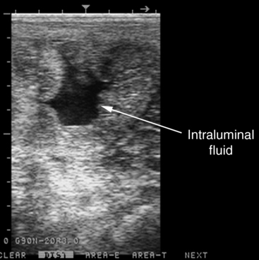
Bacterial Endometritis


The pathogenesis of bacterial endometritis in the mare is complex and has not been completely elucidated. Many mares that are not pregnant at the 14-day pregnancy check or that have early fetal loss do not have vaginal discharge. However, before and during the palpation per rectum, one should observe the vulva for exudate that might indicate uterine infection. Vaginal speculum examination may reveal variable volume of exudate at the external cervical os and on the cranial vaginal floor. Transrectal ultrasonography should always be performed to provide information about any abnormal uterine contents, especially if the mare is in estrus. The volume and echogenicity of intraluminal fluid should be noted. Detection of fluid during diestrus invariably indicates that the mare has active endometritis(Figure 169-1). Analysis of the mare’s cycling pattern may indicate shortened interestrous intervals induced by endogenous release of luteolytic prostaglandin F (PGF) from the inflamed uterus. Endometritis should be ruled out in mares that do not become pregnant after being properly managed, especially those that have distinct endometrial edema at pregnancy examination on days 12 to 14 postovulation.


Stay updated, free articles. Join our Telegram channel

May 28, 2016 | Posted by in EQUINE MEDICINE | Comments Off on Diseases of the Uterus

Full access? Get Clinical Tree

Get Clinical Tree app for offline access