Chapter 8 Diseases of the Teats and Udder
The udder of a dairy cow consists of four separate glands suspended by medial and lateral collagenous laminae. The medial laminae are more elastic, especially cranially, than the lateral laminae and are paired. Caudally, the medial laminae are more collagenous and originate from the subpelvic tendon. The lateral laminae are multiple, inserting at various levels of mammary tissue, and originate from the subpelvic tendon caudally and external oblique aponeurosis cranially. The medial and lateral laminae provide udder support that is essential for udder conformation and for solid attachment to the ventral body wall. The external pudendal artery constitutes the major blood supply to the udder; the artery courses through the inguinal canal along with the pudendal vein and lymph vessels to supply the craniolateral portion of the mammary gland. Caudally, the internal pudendal artery branches into the ventral perineal artery at the level of the ischiatic arch and courses caudally to the vulva, along the perineum to the base of the rear quarters. The caudal superficial epigastric vein (subcutaneous abdominal vein or milk vein) is the major venous return from the mammary gland. Located superficially along the ventral abdomen, it courses cranially to the “milk well” where it enters the abdomen to join the internal thoracic vein, draining first into the subclavian vein, and finally into the cranial vena cava. Lymph drainage moves dorsally and caudally to the superficial inguinal (mammary, supramammary) lymph nodes, which can be palpated by following the rear quarter dorsally until it ends, then palpating deep just above the gland along the lateral laminae. Although the prefemoral (subiliac) lymph nodes that are located in the aponeurosis of the external abdominal oblique muscle, approximately 15 cm dorsal to the patella, are not strictly drainage lymph nodes of the udder, they should be palpated as part of the routine physical examination of cattle. Because of their regional proximity and combined lymphatic drainage with the supramammary lymph nodes into the medial iliac lymph nodes, many cows with mastitis have obvious lymphadenopathy of the prefemoral lymph nodes, and they are easily palpable.
UDDER
Premature Development
Premature symmetric development of the udder in calves and heifers has been associated with chronic estrogenic stimulation resulting from cystic ovaries, feedstuffs containing excessive estrogens, and the mycotoxin zearalenone. In some cases, udder development is accompanied by vulvar swelling. When the more common etiology of ingesting feed containing estrogenic substances has occurred, multiple heifers within a group are typically affected, and successful resolution of the mammary development requires removal of the contributing feed material. Idiopathic symmetric udder development in individual heifers is occasionally encountered when no obvious endocrinologic or intoxicant cause can be elucidated. Asymmetric gland enlargement in group-reared heifers should always raise suspicion of mastitis secondary to cross-sucking.
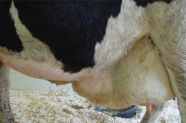
Breakdown or Loss of Support Apparatus
Breakdown of either the lateral or the medial udder supports can occur. Medial laminae breakdown causes the medial longitudinal groove between the left and right halves of the udder to disappear and causes the teats to project laterally. Loss of lateral support laminae causes the halves of the udder to project ventrally to the level of the hocks or lower. Occasionally cows lose fore udder support such that the forequarters appear detached from the ventral abdominal wall, and a hand may be inserted between the skin covering the glandular tissue and the ventral body wall. Similarly loss of rear udder attachment tends to make the rear udder pendulous without clearly defined udder attachment and obvious stretching of the skin in the escutcheon region. In the latter condition, the rear quarters no longer appear to curve up to the escutcheon but simply hang.
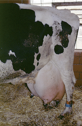
All these various deficiencies in udder support predispose to udder edema, teat and udder injuries, and mastitis. Edema is worsened by the pendulous nature of the udder, reduced venous and lymphatic return, and trauma. Injuries to the teat and udder in cows with pendulous udders (Figure 8-1) result from environmental trauma that includes contact of the udder with flooring when the cow is recumbent and direct damage from claws and dewclaws or from being stepped on by neighboring cows. Mastitis is predisposed to by environmental contamination of the teats and udder, teat injuries that affect milkout, and imperfect milkout caused by persistent edema in the floor of the udder. In some cows—especially those with severe loss of median support—it may not be possible to attach a milking machine claw simultaneously to seriously deviated teats. The result often is mastitis or culling because of milking difficulties. In addition, purebred cattle that are classified are discriminated against in classification score if these undesirable mammary characteristics are present.
Etiology of udder breakdown is complex and consists of genetic, nutritional, and management factors. Although udder breakdown is largely thought of as a problem in multiparous cows, in herds that approach an average of 25,000 lb per lactation, breakdown of the udder may occur at earlier ages.
No treatments exist for the ligament ruptures. Prevention of the condition is also problematic because other than genetic selection and control and prompt treatment of excessive parturient edema little else can be done.
Hematomas
Etiology
Self-induced trauma as a result of awkward efforts to rise or lie down and external trauma from butting or kicking by other cows are theorized as the causes of udder hematomas, but injuries from these sources seldom are confirmed. Caudal udder hematomas originating in the escutcheon region may represent thrombosis and/or rupture of the perineal vein because they tend to occur during the dry period. Udder hematomas, regardless of cause, are dangerous because blood accumulates subcutaneously, allowing massive blood loss. In addition, the exact location of the bleeding often is impossible to determine clinically because of the extensive venous plexus. Surgical attempts at finding the bleeding vessel are often futile and may lead to excessive blood loss.
Signs
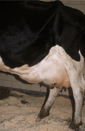
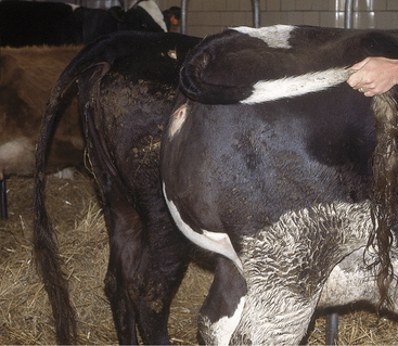
Soft tissue swellings immediately cranial to the udder are most common in lactating dairy cattle (Figure 8-2), whereas extreme swelling in the escutcheon region ventral to the vulva and dorsal to the rear quarters is more common in dry cows (Figure 8-3). The swelling may be fluctuant, soft, or firm, depending on the amount of blood causing the distention; usually it is painless and cool. Rare instances of hematomas between the base of the udder and ventral body wall also have been encountered.
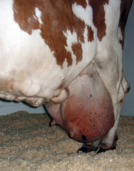
Figure 8-2 Udder hematoma apparent as soft tissue swelling cranial to the udder. The hematoma also has infiltrated the medial laminae to separate the forequarters pathologically.
Progressive enlargement of the swelling coupled with progressive anemia signal a guarded prognosis for cattle affected with udder hematomas. Signs of anemia include pallor of the mucous membranes and teats (if nonpigmented skin), elevated heart and respiratory rate, and weakness. Cattle with udder hematomas that progressively enlarge may die over 2 to 7 days.
Diagnosis
Progressive fluctuant swelling adjacent to the udder coupled with progressive anemia and absence of fever usually are sufficient for diagnosis. Ultrasound may be used to confirm the presence of a fluid-filled mass but does not always make a definitive diagnosis on its own. Ultrasonographic distinction between an abscess and a hematoma can be valuable because clinical experience suggests that aspiration of a hematoma, even under controlled and aseptic conditions, will frequently be associated with subsequent abscess formation. Ultrasonographic evidence of gas shadowing within an encapsulated mass should be taken as proof of an abscess, but mixed echogenicity images can be obtained with both abscesses and hematomas. Abscesses tend to be warm, painful, and may cause fever in the affected cow. Seromas are unusual adjacent to the udder but would give similar signs of swelling. However, seromas usually do not enlarge as much as a hematoma in this location, and progressive anemia would not be expected with a seroma.
In confusing cases, an aspirate under sterile condition may be needed to differentiate hematoma from an abscess or seroma. Clinicians should be reluctant to aspirate known hematomas for fear of introducing infection or disturbing pressure equilibrium that might allow further bleeding.
Treatment
For management of mammary gland hematomas, box stall rest and close monitoring of the animal at 12- to 24-hour intervals are important components of therapy. Complete blood counts (CBCs) and coagulation panels may be indicated to rule out bleeding disorders. Flunixin and other nonsteroidal antiinflammatory drugs (NSAIDs) should be withheld during the period of active hemorrhage.
In general, bleeding disorders of cattle are rare and are unlikely causes of udder hematomas. Occasionally consumptive thrombocytopenia may occur in cows with udder hematomas. Cows that experience hemorrhage sufficient to reduce packed cell volumes (PCVs) to 10% to 12% and have a heart rate exceeding 100 beats/min and a respiratory rate in excess of 60 breaths/min require a whole blood transfusion of 5 to 8 L. Platelet-rich whole blood from a healthy donor (negative for bovine leukemia virus [BLV], bovine virus diarrhea virus [BVDV], bluetongue virus, anaplasmosis, and Johne’s disease) is important if anemia becomes severe.
Stabilization of the size of the hematoma and other clinical signs are positive prognostic indicators, whereas progressive anemia and enlargement of the hematoma despite therapy are negative indicators. Affected cows should be separated from herdmates to avoid further trauma. Incision of udder hematomas to arrest bleeding is unrewarding, may exacerbate bleeding, and therefore is contraindicated. Stabilized udder hematomas eventually resorb, but some may abscess and drain by 4 weeks because of pressure necrosis of overlying skin. When drainage occurs, large necrotic clots of blood and serosanguineous fluid drain from the area. Surgical debridement of naturally draining hematomas is not indicated except in chronic cases (.4 weeks) with abscessation, in which case ultrasound guidance should be considered. The condition does not recur once fully resolved. Udder asymmetry, abnormal teat deviation, and persistent residual edema are common sequelae to udder hematoma and abscess resolution, and although these are of limited economic impact in a grade cow, they may be a considerable frustration for the owners of show and pedigree cows.
Abscesses
Etiology
Udder abscesses may appear anywhere in the mammary tissue or adjacent to the glands. Frequently skin puncture with subsequent abscessation is suspected when obvious abscesses appear in quarters having completely normal secretion and no evidence of mastitis. Endogenous abscesses can form secondary to mastitis with abscessation, as is typical of mastitis caused by Arcanobacterium pyogenes. However, this discussion will be confined to udder abscesses requiring percutaneous drainage, and discussion of mastitis origin abscesses will be addressed in the mastitis section.
Most udder abscesses harbor typical contaminants such as A. pyogenes or staphylococci, but occasionally other organisms may be involved.
Abscesses appear as firm, warm swellings that may be either distinct or indistinct from gland parenchyma (Figure 8-4). Palpation of the swelling may be painful to the affected cow. Milk from the affected quarter usually is normal, and the abscesses tend to be well-encapsulated.
Diagnosis
Physical signs usually are sufficient for diagnosis, but ultrasonography or aspiration may be indicated if the owner is impatient regarding diagnosis and therapy. Ultrasound may be beneficial to diagnosis and treatment of udder abscesses because flocculent material may be observed, and major vessels overlying the lesion may be located if aspiration and drainage are necessary. A thick capsule around the abscess is usually observed.
Treatment
A conservative approach usually is rewarded by eventual natural rupture and drainage of the abscesses in 2 to 8 weeks. This has been standard treatment because practitioners fear lancing anything in the udder because of the extensive blood supply to the entire organ. Conservative treatment probably still is the safest. The only risk from conservative therapy is the same for neglected abscessation in other tissues—that is, chronic antigenic stimuli that may predispose to glomerulonephritis, amyloidosis, or bacteremia, which may cause endocarditis or other infectious disease.
Therefore the practitioner may be pressured by owners of valuable or show cattle to encourage early drainage. Ultrasound and aspiration are indicated before draining, as discussed previously.
Following natural or surgical drainage, the abscess cavity should be flushed daily with dilute antiseptics or saline, and the drainage hole should be kept open, lest premature closure allow the abscess to reform. Usually antibiotic treatments are unnecessary.
Thrombophlebitis and Abscessation of the Milk Vein.
Thrombophlebitis of the mammary vein is an occasional complication of venipuncture at this site (Figure 8-5). This vessel seems attractive for the intravenous (IV) administration of pharmaceuticals, particularly in pit parlors and for producers for whom its size and accessibility make it a less challenging alternative compared with the jugular or coccygeal veins. However, the consequences of mammary thrombophlebitis with respect to udder symmetry and future production are sinister enough that it should never be used in show or valuable individual cows. It should only be used under considerable duress even in grade cattle. Abscesses may develop secondary to phlebitis from the use of contaminated needles or subsequent to hematoma formation when vascular damage and perivascular leakage occur as the cow resists the procedure. As with all cases of thrombophlebitis there is a risk of embolic spread, potentially causing endocarditis or nephritis. If treatment is initiated immediately following the inciting attempted venipuncture, antiinflammatory and antimicrobial therapy is indicated. However, veterinary attention is often only sought after abscessation has already occurred, at which time the goal of therapy should be surgical drainage followed by antimicrobial therapy. It can be challenging to avoid significant blood loss when lancing such abscesses because of the highly vascular nature of the region, and ideally the procedure should be performed under ultrasound guidance. The bacterial species implicated include the common pyogenic anaerobes, and antimicrobial therapy should include beta-lactam antibiotics. Treatment of valuable cattle may include rifampin under appropriate extra-label drug use guidelines.
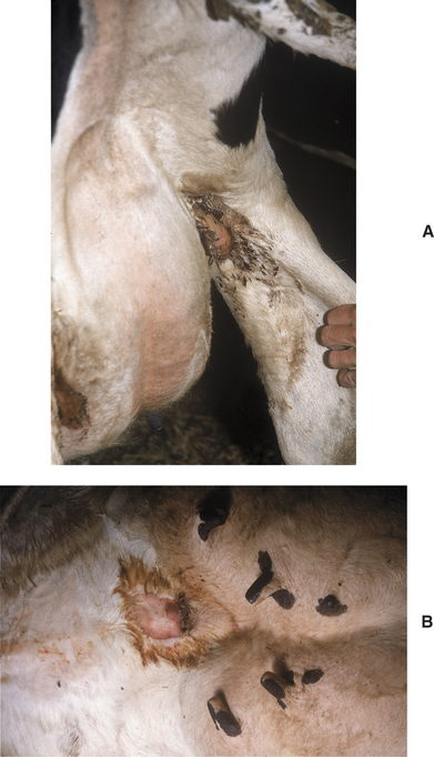
Udder Cleft Dermatitis
Etiology
Udder sores are foul-smelling areas of moist dermatitis that result from pressure necrosis of skin associated with periparturient udder engorgement and edema. Common locations include the skin reflection between the medial thigh and dorsal attachment of the lateral udder, on the ventral midline immediately adjacent to the median septum of the foreudder, and on the median septum of the udder—either between the forequarters or in the fold that is centered between the four quarters.
Pressure necrosis associated with udder edema is enhanced by frictional injury and chafing with limb and udder movement. The abraded skin oozes serum, which, coupled with the omnipresent skin hair, leads to moist dermatitis. Finally, opportunistic anaerobic bacteria such as Fusobacterium necrophorum and A. pyogenes invade and propagate under crusts, scabs, and necrotic skin. The organisms cause the smell that distresses milkers each time they get close to the udder—hence the name “udder rot.” Chorioptic mange mites and Malassezia spp. have been incriminated in some cases.
Signs
A fetid odor similar to that found in septic metritis or retained placenta emanates from areas of moist dermatitis in the groin area or more commonly the ventral median area of the udder. Skin necrosis may be mild or severe. In the worst cases, large patches of skin (10 to 30 cm in length) may be peeled off. Matted hair, scabs, and necrotic skin are present (Figure 8-6, A and B). Myiasis may occur in warm weather. In some first-calf heifers, groin infections can be so severe that lameness may occur.
Diagnosis
The combination of necrotizing fold dermatitis and malodorous discharge in a postpartum cow is sufficient for the diagnosis of udder fold dermatitis.
Treatment
Although the principles of treatment are straightforward, client compliance may be lacking because of time constraints, and udder cleft dermatitis is not treated on larger dairies.
For dairies that request therapy, a commercially available topically applied wound spray (Granulex Aerosol Spray, Pfizer Inc., Exton, PA) consisting of a mixture of castor oil, balsam of Peru, and trypsin has been used successfully by field veterinarians for the treatment of udder cleft dermatitis. The spray is marketed for administration in humans and has been effective for treatment of decubitus in bedridden patients. Other compounded remedies have been recommended for the treatment of udder cleft dermatitis, but given the current drug compounding regulations of the U.S. Food and Drug Administration (FDA), these formulations cannot be recommended for use in food-producing animals. Patients that have developed cleft fold pyodermas secondarily to udder edema should be treated with diuretics (furosemide, 1 mg/kg) to reduce edema and anaerobiosis in the skin of the mammary gland. Diuretics are calciuretic and kaluretic, so during diuretic treatment of postpartum cows, calcium status should be monitored, and if the cow shows weakness, lassitude, and cold extremities, she should be given IV calcium gluconate and 100 g of potassium chloride orally.
The prognosis in cases of udder cleft pyoderma is excellent, but healing time may be prolonged for months in untreated cases. If the inguinal lesions are causing severe lameness, surgical debridement can speed healing.
Udder Dermatitis
Etiology
Udder dermatitis may be associated with a multitude of chemical, physical, and microbiologic causes.
Chemical causes of udder dermatitis include irritants in bedding such as hydrated lime, ammonia from urine, copper sulfate, or formaldehyde from foot baths.
Physical causes of udder skin inflammation include sunburn, frostbite, and pressure necrosis caused by decubitus. Lesions of photosensitization may appear on the sun-exposed, nonpigmented skin of teats and the udder.
Staphylococci and streptococci occasionally cause a diffuse miliary folliculitis or pustular dermatitis named “udder impetigo.” Rarely Dermatophilus congolensis affects the skin of the udder, but this tends to occur as part of a severe generalized infection. Disseminated infections of Trichophyton verrucosum may affect the skin of the udder. Viral lesions of the skin of the udder include herpes mammillitis (see the discussion of Teat Skin Infections), BVDV, bovine bluetongue virus infection, and malignant catarrhal fever (MCF).
Signs
Signs vary with specific etiology of the lesions. Chemical and physical teat skin dermatitis is characterized by extreme erythema, swelling, and evidence of pain. Vesicles may be present in extreme cases associated with irritant chemicals. The skin of the teats and the udder may be involved. Serum oozing and slight matting of the hair may be apparent.
Sunburned teat skin of dairy cattle has a similar appearance to that found in other species, and the cow may show evidence of sunburn in other locations. Affected skin is warm, painful, and may have vesicles or bullae caused by burns on nonhaired skin near the teat-udder junction. Multiple cows in the herd may show signs simultaneously and resent milking procedures because of painfully burned skin. Signs may be present on only one side of the udder if the cow preferentially lies on one side. Frostbite occurs during extreme winter cold—mostly in free stall barns and mostly in periparturient cows with udder edema that already compromises tissue circulation. Patchy areas of skin on the teats and udder become cool, discolored, swollen, and then turn leathery and completely cold. Frostbite must be differentiated from herpes mammillitis.
Other physical causes of udder dermatitis include photosensitization, which again would be apparent because of signs in other depigmented regions of skin and known exposure to sunshine.
Pressure necrosis or decubital sores from extended periods of recumbency occur mostly in cattle with very pendulous udders but may also occur in downer cattle or lame cows that lie down more than normal. Such sores often are located where the medial hock makes contact with the udder. Lesions initially are reddened, ooze serum, and then slough, leaving a necrotic crater-like lesion in the udder.
The clinical signs of infectious dermatitis vary with the causative agent. Staphylococcal dermatitis causes a diffuse folliculitis with small raised tufts of hair joined with dry or moist exudate. Pustules may be apparent in the worst cases. Usually only one or a few cows in the herd are affected, but occasionally outbreaks of pustular dermatitis have been observed. D. congolensis may appear as larger confluent areas of folliculitis with dry or moist crusts that hold tufts of hair together. Plucking these tufts of hair or crusts may reveal purulent material on the underside of the crust or adjacent skin. Cows with dermatophilosis on the skin of the udder usually have other obvious Dermatophilus lesions.
T. verrucosum lesions on the udder are circular or patchy alopecic areas that are 1.0 to 10.0 cm in diameter. Most lesions have crusts as observed in ringworm lesions in other locations, but some lesions may appear as moist alopecic regions as a result of the paucity of hair in certain regions of the udder. Other areas of ringworm infection usually are identified during inspection of the cow. Herpes mammillitis lesions coexist on the skin of the teats and udder. Recognition of bullae or vesicles in early cases is imperative to diagnosis.
Herpes mammillitis (bovine herpes virus 2 [BHV2]) most often occurs in first-calf heifers, and usually more than one animal will be affected. Another herpesvirus, BHV4, the DN599 strain of BHV that has been isolated from the respiratory tract of cattle, may be capable of causing pustular mammary dermatitis. Multiple vesicles and pustules from 1.0 to 10.0 mm in diameter have been observed on the udder of lactating cows, and lesions have been observed in farm workers.
Treatment
Treatment for chemical dermatitis only requires gentle washing of the udder with warm water and removal of the offending agent from the cow’s skin. Individual cows may be sensitive to a chemical despite the majority of cows in the herd being exposed to the same chemical yet remaining unaffected. Warm water cleansing of the udder to remove residual chemical followed by application of aloe or lanolin products is recommended.
Physical causes of dermatitis are best treated by preventing further exposure to the specific physical cause and by symptomatic therapy. Sunburned cows must be kept inside or provided shade if at pasture. Cool water compresses followed by aloe or lanolin ointments to deter skin cracking and peeling caused by dryness are helpful. Udder supports may help prevent sunburn. Frostbite is best prevented by therapy for excessive udder edema and keeping periparturient cows well bedded for warmth. Periparturient cows in free stalls during extreme cold are at greatest risk because free stall beds usually are poorly bedded for warmth. Once frostbite has occurred, careful sharp debridement of necrotic tissue and protection against further injury are the only potentially effective treatments.
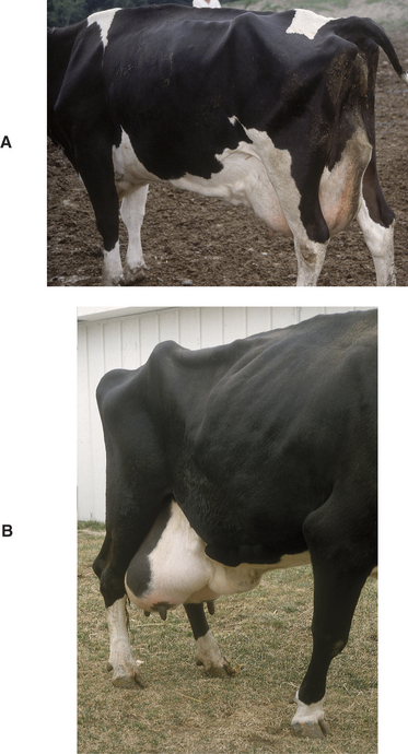
Pressure necrosis or decubital sores are treated by providing soft bedding for cows that spend more time than normal recumbent and minimizing udder edema. Extremely pendulous udders are at greater risk for decubital sores. Treatment of decubital sores requires gentle cleansing and debridement following by the application of Granulex spray. Decubital sores may require weeks or months to heal.
Photosensitization must be treated by avoiding further sunlight, applying cleansing lesions, debriding necrotic skin, and determining the cause of photosensitization.
Microbiologic causes of udder skin dermatitis are managed according to the specific cause. Staphylococcal folliculitis is treated by clipping the hair on the udder, washing gently with povidone iodine scrub solutions, rinsing with water, and drying. Washing and rinsing should be done once or twice daily. Antibiotics generally are not necessary. Filthy environmental contributing factors should be eliminated.
D. congolensis infections should be treated by clipping the hair of the udder followed by removal of all crusts through gentle washing with povidone iodine scrubs and drying. In those with severe or generalized dermatophilosis, penicillin (22,000 IU/kg body weight, once or twice daily, intramuscularly [IM]) may be necessary for 5 to 7 days in addition to local therapy.
Ringworm lesions can be managed by clipping the hair on the udder (especially if it is long or filthy) followed by topical application of chlorine bleach diluted 50:50 with water, miconazole, or clotrimazole ointments to the lesions once or twice daily. Care should be taken such that antifungal medications do not contaminate milk during milking procedures.
No specific treatment exists for udder lesions resulting from herpes mammillitis or other dermatopathic viruses.
Edema
Etiology
Udder edema, also known as “cake,” may be physiologic or pathologic. Physiologic udder edema begins several weeks before calving and is more prominent in heifers preparing to have their first calf. Many questions remain unanswered as regards the etiology of udder edema. Genetic factors certainly exist, and bull stud services sometimes grade production sires by probability of udder edema in their female offspring. Individual cows with severe or pathologic edema should be examined to rule out medical considerations that could contribute to ventral or udder edema. Some conditions to consider include cardiac conditions, caudal vena caval thrombosis, mammary vein thrombosis, and hypoproteinemia resulting from one of a number of diseases. Physical examination and serum chemistry screens may be helpful in the evaluation of such individuals.
Postparturient metritis has been associated with persistence of physiologic or pathologic udder edema by some owners and veterinarians. The pathophysiology of this relationship is unknown.
When many cows in a herd have either severe physiologic udder edema or pathologic udder edema, herd-based causes must be considered. Although feeding excessive grain to dry cows and early lactation cows has long been discussed as a cause of such endemic udder edema problems, feeding trials do not support this theory. Similarly, high protein diets do not seem to be directly involved. Currently, excessive total dietary potassium and sodium are considered possible culprits in herd-wide udder edema problems. Total intake of potassium may be excessive in some instances when high quality alfalfa haylage constitutes a major portion of the ration. Forages harvested from land that is fertilized repeatedly with manure are becoming an increasing problem because of their high potassium content. One article recommends no more than 227 g/day/head of potassium in heifers. Similarly sodium levels may be excessive when considering total available sodium in the basic ration, water, and mineral additives plus or minus free choice salt.
Hypoproteinemia and especially low albumin fractions in affected cows also may contribute to udder edema. Metabolic profiles need to be performed to assess this possibility and determine the origin.
Signs
Physiologic udder edema may start in the rear udder, fore udder, in the left or right half of the udder, or symmetrically in all four quarters. Edema tends to be most prominent in the rear quarters and floor of the udder (Figure 8-7). Cows with moderate to severe udder edema usually have a variable degree of ventral edema extending from the fore udder toward the brisket.

Figure 8-7 Forced abduction of the hind limbs is obvious because of severe udder edema in this first-calf heifer.
Udder edema pits and associated ventral edema may be soft and fluctuant or firm and pitting. Physiologic edema increases up to calving and then begins to gradually resolve over 2 to 4 weeks.
Pathologic edema persists longer than physiologic edema. Pathologic edema may be present for months following parturition or for the entire lactation. The tendency for pathologic edema is increased in cows with breakdown of udder support structures, and conversely pathologic udder edema may contribute to breakdown of udder support structures. Therefore severe edema may affect a cow’s longevity and classification in some instances.
Pronounced udder edema interferes with complete milkout because it causes the affected cow discomfort, and milking may accentuate that discomfort. In addition, interstitial edema in the mammary glands may cause pressure differentials that interfere with normal production and let down of milk. Therefore chronic or pathologic edema may have a negative effect on the lactation potential because cattle never reach their projected production. Interference with complete milkout resulting from pain, as well as mechanical or pressure influences, also may lead to postmilking leakage of milk in cows with severe udder edema. This translates into an increased risk of mastitis.
Cows with udder edema do not act ill but may be uncomfortable or painful because of the swollen, edematous udder swinging as they move or from constantly being irritated by limb movement as they walk. In addition, when resting, the cow may tend to lie in lateral recumbency with the hind limbs extended to reduce body pressure on the udder.
Diagnosis
Diagnosis is based on inspection, palpation, evaluation of milk secretions to rule out mastitis, and ruling out conditions such as udder abscess or hematoma. Pitting edema should be present, especially on the floor of the udder. Pitting edema may be evident over the entire udder in severe cases, and ventral edema frequently coexists in these instances.
Treatment
Treatment of individual preparturient or postparturient cows is indicated when edema has the potential to break down the udder support structure. Treatment also is indicated for preparturient cows having severe udder edema associated with leakage of milk from one or more teats.
Diuretics constitute the principal treatments for udder edema. Preparturient treatment of cattle with furosemide (0.5 to 1.0 mg/kg body weight) as an initial treatment followed by decreasing dosages of furosemide once or twice daily for 2 to 4 days is commonly used. Salt restriction should be considered. Premilking may be indicated in preparturient cows with severe udder edema that are leaking milk. This must be an individual decision based on the owner’s experience with premilking. Obviously if the option of premilking is selected, the newborn calf will require colostrum from another cow. Although premilking is controversial, some owners of show cattle swear by the technique to preserve udder conformation. Udder supports also may be helpful if fitted properly.
A word of warning about furosemide—urinary losses of calcium may be sufficient to increase the risk of periparturient hypocalcemia, and this should be anticipated in multiparous cows receiving multiple doses of the drug.
Parturient and postparturient cows judged to need treatment for udder edema may receive either furosemide or dexamethasone-diuretic combinations orally.
Individual cows may respond to one product better than the other, but this is impossible to predict. Furosemide seems to work well in some herds, whereas the dexamethasone-diuretic combination is superior in others. When considering dexamethasone-diuretic combinations, the veterinarian should first rule out contraindications to corticosteroid use. Udder supports and salt restriction may or may not be practical but should be considered. Nursing procedures including udder massage, more frequent milking, and mild exercise are helpful but labor intensive. Metritis should be ruled out or treated.
In herds with endemic udder edema, nutritional consultations are imperative to evaluate anion-cation balance. Total potassium, total sodium, and serum chemistry to profile affected and nonaffected cows should be performed. Diets with anionic salt supplementation and those with added antioxidants may show some tendency to diminish udder edema in affected herds. Water and availability of free choice salt or salt-mineral combinations should be included in the nutritional evaluation.
Hemorrhage into a Gland
Etiology
Hemorrhage into one or more glands is common at parturition in cows with severe udder edema or pendulous udders that have been traumatized by hind limb movement and awkward posture during recumbency. Milk from one or more quarters contains blood and may appear as pink, red, or reddish-brown with blood clots. Generally this condition clears within four to eight milkings and is not a major problem. Cows with bloody milk should be watched closely for mastitis because blood provides an excellent growth medium for bacteria.
As opposed to the usually innocuous parturient hemorrhage described previously, severe hemorrhage involving one or more quarters occasionally is observed in dairy cattle during lactation. The cause is unknown, but nonspecific trauma usually is suspected. Thrombocytopenia has been confirmed in some but not all of these cows, and when it is identified, it is not known whether it was as cause or effect.
Signs
The chief complaint for a cow with intramammary hemorrhage is persistent blood-stained milk from one or more quarters. Anemia may develop if extensive bleeding continues to occur over several milkings. The milk usually is red rather than pink, and blood clots are obvious. Large intraluminal clots occasionally plug the papillary duct, causing difficulty in milkout.
Diagnosis
The clinical signs of intramammary hemorrhage are sufficiently diagnostic, but laboratory work should be performed to assess thrombocyte numbers. Coagulation profiles that may be used to incriminate specific bleeding disorders are frequently unreliable in cattle. Specific causes of intramammary hemorrhage are seldom identified. Intramammary hemorrhage may increase the risk for mastitis.
Treatment
Decisions for appropriate therapy are difficult because of the likelihood of iatrogenic complications. An apparently obvious solution is to stop milking the affected quarters—thereby stopping further blood loss and allowing pressure to build up in the gland to deter further bleeding. However, this approach may provoke such severe blood clotting in the ductules, gland cistern, and teat cistern that future milking is impossible. On the other hand, once- or twice-a-day milking usually allows blood clots to be stripped out but causes more blood loss and may allow continued bleeding from whatever vessel is leaking. Generally reduction of milking frequency (usually to once daily) has been considered to be an optimal management for intramammary hemorrhage. Blood clots that form may be stripped out as they form and do not ruin future potential. If this approach does not resolve the problem within several days, a decision to stop milking and risk severe cisternal clots must be considered to save the cow.
Thrombocytopenia and other coagulation defects should be excluded as causes of the hemorrhage. When thrombocytopenia or severe anemia (PCV, 10%) exists, an immediate fresh whole blood transfusion from a donor that is uninfected by BLV, BVDV, bluetongue virus, anaplasmosis, and Johne’s disease should be administered. Approximately 4 to 6 L of blood should be transfused at a single time. Blood transfusion also may become necessary regardless of cause if the cow’s anemia becomes severe enough to warrant transfusion.
MAMMARY TUMORS
Except for fibropapillomas (warts), mammary tumors in dairy cattle are rare. Lymphosarcoma is the most common and will be considered below under a separate heading. Relatively few dairy cows live to an old age, but those that do still have a very low incidence of mammary tumors. Both squamous cell carcinomas and mammary gland adenocarcinomas have been observed in older dairy cattle ($15 years). Fibropapillomas (warts) are more common on the skin of the teats but also may appear on the skin of the udder. Many other sporadic tumors, including fibromas, fibrosarcomas, and papillary adenomas, have been reported.
Lymphosarcoma
Lymphosarcoma is the most common tumor to appear within the gland and associated lymph nodes in dairy cattle. Focal and diffuse infiltration of the gland with lymphosarcoma and rarely adenocarcinoma has been observed (Figure 8-8, A and B). The mammary gland is hardly ever the only site of lymphoma infiltration, however. Usually tumor masses in other target organs or lymph nodes supersede mammary involvement. Affected glands may merely appear edematous rather than firm, and secretions may appear normal. Diffuse lymphocytic infiltration of the udder may appear similar to the diffuse mild edema that develops in hypoproteinemic cattle. The mammary lymph nodes (superficial inguinal) may be enlarged because of lymphosarcoma or chronic inflammation and should routinely be palpated during physical examination.

Figure 8-8 A, A mature Holstein affected with lymphosarcoma. The right supramammary lymph node is markedly enlarged and appears as a firm swelling in the dorsal aspect of the right rear mammary gland. The cow also had diffuse infiltration of the mammary gland and cardiac neoplasia. B, A 14-year-old Holstein with mammary gland adenocarcinoma. There was diffuse neoplastic involvement of the gland.
(Photo courtesy R.H. Whitlock.)
Juvenile tumors of the mammary gland have been observed in two heifers referred to our clinic. Surgical extirpation of these masses was performed before udder development. Juvenile tumors found to be fibromas associated with the teat also have been recognized in two yearling heifers.
Signs
Affected cows have a varied clinical presentation ranging from focal enlargement to diffuse and massive udder enlargement. Some squamous cell carcinomas are ulcerated, firm, and pink with a malodorous smell. Precursor papilloma or epithelioma masses may have been observed by the owner. Fibropapillomas or warts are obvious and are of little concern.
Juvenile tumors of the gland cause an obvious enlargement that may be confused with mastitis in the undeveloped udder.
Diagnosis
Biopsy or aspirate of a suspicious mass is essential for diagnosis. Biopsy of the gland may be performed if diffuse lymphosarcoma is suspected. Commercial true-cut biopsy needles work very well for mammary gland biopsies, and the procedure is safe.
Treatment
Juvenile tumors of the gland may be excised, but prognosis for production in the affected gland must be guarded. However, in the two juvenile gland tumors observed by Dr. Rebhun and in the two reported teat skin fibromas in yearlings, long-term follow-up evaluation indicated that all glands that had undergone surgery were functional at first calving. Squamous cell carcinomas may be treated according to severity. Cryosurgery may suffice in early stages of the disease, but udder amputation may be required in advanced cases. Treatment for lymphosarcoma is rare and generally of limited success.
Udder Amputation
Hemimastectomy or radical mastectomy is rarely performed in cattle. Indications for this in valuable or pet cattle would be neoplasia, chronic incurable mastitis, and/or suspensory apparatus failure that causes the udder to sag and be persistently traumatized (Figure 8-9). Individual animals that undergo udder amputation must be in good general condition. Significant blood loss can occur with udder amputation because of the vascularity of the mammary gland. Cattle with septic mastitis or in poor physical condition should not undergo udder amputation until their physical condition is significantly improved.
The cow must be placed in dorsal recumbency and preferably under general anesthesia. A fusiform skin incision is made to facilitate subsequent skin closure. Therefore the lateral incisions must extend to the junction between the middle third and dorsal third of the udder to allow sufficient skin for closure under minimal tension. Following skin incision, the dissection is first directed toward the inguinal canal where the pudendal arteries and then vein are ligated. The procedure is repeated on the contralateral side. Using curved Mayo scissors, the loose fascia on the proximal aspect of both lateral laminae is incised starting cranially and extending caudally until the left and right perineal arteries and veins are located and double ligated. To minimize systemic blood loss (through retention in the mammary gland), the caudal superficial epigastric veins are ligated last. The lateral laminae are then sharply transected and dissection extended on the dorsal aspect of the mammary gland to complete the excision.
Before closure, one should attempt to control the extent of postoperative seromas by placing two, 2.5-cm Penrose drains in the space between the ventral abdominal fascia and the subcutaneous tissues on either side of the midline along the ventral abdomen. Stab incisions are used to create portals for the drains to exit on either side of the incision and secured to the skin using a single interrupted suture to avoid accidental drain removal.
Closure is performed in three layers. The subcutaneous tissue is closed in two layers using no. 2 absorbable suture material (such as chromic catgut or polyglactin 910) in a simple continuous pattern. Every 2 or 3 cm, the subcutaneous sutures should penetrate the abdominal fascia to reduce dead space. The skin is closed in a forward interlocking pattern with a nonabsorbable material (such as polyamide), and a stent is sutured over the incision to help diminish the tension on the incision.
TEAT DISEASE
The tissue layers in the teats of dairy cattle include skin, inner fibrous, stroma, and mucosa. Stroma is a vascular and muscular layer containing veins that drain to the large subcutaneous venous plexus at the juncture of teat and udder. The inner fibrous layer is a thin membrane that is interposed between the mucosa and the stroma (Figure 8-10).

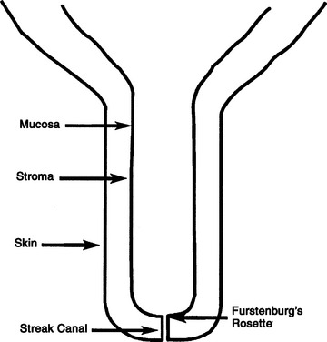
Figure 8-10 Schematic of the bovine teat to illustrate the basic anatomic terms used in this chapter.
The papillary duct (teat canal) or streak canal is the exit for milk from the teat sinus or cistern and is surrounded by the teat sphincter muscle. The papillary duct and sphincter muscle represent a significant component of the defense mechanism against mastitis, and they are the most frequently injured portions of the teat.
The streak canal in the healthy state acts both as a valvular obstruction to milk flow and as a unique deterrent to ascending infection of the gland. The canal is lined with keratin as a result of a specialized stratified squamous epithelium arranged longitudinally. Keratin in the papillary duct binds bacteria and then desquamates to form a plug with antimicrobial activities that may deter bacterial entrance.
Congenital Anomalies
Etiology and Signs
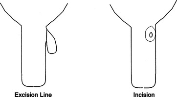
Supernumerary teats are the most common congenital abnormality, which is likely heritable in dairy cattle. There has been little genetic selection away from this trait in heifers simply because it is not reported back to artificial insemination stud services and because the problem is so easily treated. Supernumerary teats are extremely common in certain lines of cattle—especially Guernseys. Usually one to four extra teats is observed, although more can occur. Generally the supernumerary teats are placed caudal to the rear quarter teats or between the rear and forequarter teats. These “extra” teats require treatment, lest functional glands evolve that would likely become infected because they cannot be milked effectively. Such infections provide a chronic source of infection for other quarters in the herd.
Supernumerary teats that are joined to one of the four major teats have been called webbed or “Siamese” teats. These may appear as distinct teats or only as small raised areas on the wall of one of the major teats (Figure 8-11, A to D). Although these teats frequently have a separate gland of their own, they may communicate with the teat or gland cistern of the major teat (Figure 8-12). In either case, these joined teats require special treatment and careful differentiation from simple supernumerary teats, lest future production of the gland be compromised.

Figure 8-11 A, Webbed teats may appear as a distinct teat. B, An enlarged proximal portion of teat. C, Only as a small tissue mass on a normal-sized teat. D, Or as clusters.

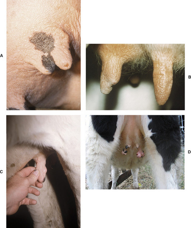
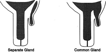
Figure 8-12 Co-joined supernumerary teats may communicate with a separate gland or the gland of the major teat.
Keratinized corns or keratomas on the teats of heifers have been recognized. These structures are tightly adherent to the teat end, grow progressively, and their physical weight stretches and elongates the affected teat (Figure 8-13).
Juvenile tumors of the skin adjacent to the teats of two heifers have been described and were found to be a fibroma and a fibrosarcoma.
Diagnosis
Diagnosis of supernumerary teats, Siamese teats, and keratinized corns simply requires inspection. Supernumerary teats cojoined to a major teat require more careful consideration and treatment, but if identified in calfhood, the treatment is the same. Tumors associated with the teat require biopsy to allow definitive diagnosis.
Treatment
Heifer calves should be examined for the presence of supernumerary teats at a routine time during the first 8 months of life. Most veterinarians perform this examination when vaccinating 4- to 8-month-old calves for brucellosis. Following restraint of the heifer, the supernumerary teats are grasped and cut off with scissors at the point where the skin of the teat meets the skin of the udder. A suitable antiseptic is applied to the wound following removal, but sutures generally are not used except when a cosmetic appearance is required immediately. Care must be exercised when removing supernumerary teats, lest a true teat be removed accidentally.
If confusion exists as to which teats are the true ones, the heifer should be allowed to grow for a few months and then be rechecked. It is important, however, to force owners to check for supernumerary teats when the heifers are young so that surgical removal is easier for both the animal and the veterinarian. Removing supernumerary teats on a 2-year-old heifer that is already springing can be difficult and unpleasant for all involved parties.
Supernumerary teats cojoined to a major teat (webbed teats) need to be repaired surgically rather than just snipped off. The repair should be performed when the teat is large enough to be manipulated easily and then sutured. Many surgeons prefer to operate on animals at approximately 8 months of age. Aseptic technique is essential because infection of the future mammary gland is a major risk. Following an 8- to 12-hour fast, the heifer is sedated with xylazine (4 to 5 mg/kg/100 lb body weight, IV), tied in dorsal recumbency, and the hair is clipped from the udder near the extra teat. Following routine preparation, a fenestrated drape is placed, and the supernumerary teat is excised by scalpel or scissors. This excision is nearly flush with the skin of the main teat but should leave enough skin to close the wound (Figure 8-14). After excision, the mucosa of the rudimentary teat should be closed with fine synthetic absorbable suture (e.g., Monocryl, Ethicon, Johnson & Johnson, Somerville, NJ), such as 3-0 or 4-0. The skin and stroma are sutured as a single layer using interrupted vertical mattress sutures or alternatively closed with individual layers. Many suture materials are suitable for stroma and skin closure, but absorbable sutures such as 3-0 or 4-0 Vicryl (Ethicon, Johnson & Johnson, Somerville, NJ) or Monocryl are popular because they do not require subsequent removal and associated restraint. Antibiotics such as IM penicillin (22,000 IU/kg) should be administered preoperatively and for 2 to 3 days postoperatively.

Figure 8-14 Schematic illustration of excision line for co-joined super numerary teat (left) and outline of a resultant incision (right). The inner circle in the incision represents mucosa, and the outer circle indicates skin.
When surgery is done at this early age, it does not matter whether the ancillary teat has a separate gland. If, however, surgery is delayed until after the first lactation, surgical repairs may become considerably more complicated because of the need to connect separate gland cisterns to the major teat.
Keratin corns or keratomas may be surgically dissected from the teat ends, but simpler means of therapy exist. A light teat bandage held on the teat by adhesive tape may be used to moisten the keratinized material, and the keratinized material may be gently separated from the teat itself after several days of soaking. Saline, lanolin and aloe mixtures, and ichthammol ointment have all been used successfully to soften keratin corns and allow subsequent removal.
Successful removal of tumors associated with the teat has been described. Future production was not destroyed in these animals; nonetheless a guarded prognosis is justified.
Teat-End Injuries
Etiology
Teat ends are the most common site of mammary injury in dairy cattle and are the most common reason for owners to seek veterinary consultation regarding the teats of dairy cattle. Teat-end injuries may affect the sphincter muscle, the streak canal, or both. Injuries to the teat end are caused by the digit or medial dewclaw of the ipsilateral limb of the affected cow or by injury from neighboring cows stepping on the teat. Teat-end injuries are more common in cows with pendulous udders or in those that have lost support laminae. Acute injuries cause inflammation, hemorrhage, and edema within the distal teat stroma and sphincter muscle. Subsequent soft tissue swelling in the teat end mechanically interferes with proper milk release from the streak canal. In addition, the streak canal epithelium and keratin may be disrupted, crushed, lacerated, partially inverted into the teat cistern, or partially inverted from the teat end. Occasional distal membranous obstruction occurs as a result of teat injuries followed by local fibrosis (Figure 8-15). Obvious laceration of the distal teat skin may be present but frequently is not. When present, lacerations tend to be at the teat end. Degloving injuries to the teat end are also occasionally encountered subsequent to claw or limb trauma when the teat becomes trapped against solid flooring. Repeated or chronic teat-end injury leads to fibrosis of the affected tissues, granulation tissue at the site of any mucosal or streak canal injury, and continued problems with milkout. Subclinical teat-end injury has been associated with defective milking machine functions such as increased vacuum pressures or overmilking.

Figure 8-15 Sonogram of the distal aspect of the teat made with a linear 12.5-MHz probe (distal to the left). A focal occlusion at the distal aspect of the teat cistern is identified as a 3-mm band of tissue. The rosette of Furstenberg and the streak canal (thin hyperechoic line at the tip of the teat) are normal.
(Courtesy Dr. Amy Yeager, Cornell University.)
In addition to traumatic injuries, teat-end ulceration is a common problem that may involve individual cows or be endemic in certain herds. Crater-like ulcers filled with dried exudate and scabs make milkout very difficult and predispose to mastitis. Many causes, including irritation from teat dips, excessive vacuum pressure, and mechanical abrasions have been suggested, but the exact cause of the lesion often is difficult to ascertain.
Signs
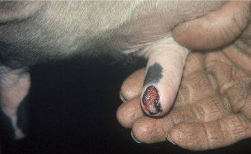
Painful soft tissue swelling of the distal teat is the cardinal sign of acute teat-end injury. The skin may be hyperemic or bruised (Figure 8-16). The cow resents any handling or manipulation of the teat end and objects to being milked. A combination of mechanical interference with milkout and pain-induced reluctance to let down milk predisposes to incomplete milkout from affected quarters. Mastitis is the feared and frequent sequela to incomplete milkout in cows with teat-end injuries. The cow is further predisposed to mastitis if the physical defense mechanism of the streak canal is compromised.

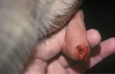
Figure 8-16 Acute teat-end injury showing diffuse swelling and a blood clot extruding from the streak canal.
Chronic teat-end injury often includes a history of acute injury followed by continued difficulty in milking. Palpation of the teat end allows detection of fibrosis in the sphincter muscle or granulation tissue dorsal to the streak canal and sphincter muscle at the ventral-most portion of the teat cistern. Pain is not as apparent with chronic teat-end injuries as in acute cases.
Treatment
Treatment of acute teat-end injuries should address both the injury and any management factors that might lead to further injury, such as overcrowding, lack of bedding, and milking machine problems.
Treatment considerations must be acceptable and logical to the milkers because the milkers are responsible for any ongoing treatment. Milkers also are subject to the end results of the cow’s pain caused by manipulation of the acutely injured teat. Unless one has milked cows, it may not be apparent exactly how difficult it is to remain patient when being kicked at by cattle that object to having injured teats handled. Client compliance necessitates empathy for the patient, as well as the people responsible for milking the cow. Advice regarding patient restraint, minimizing pain, and preventing mastitis must be included in any treatment regimen.
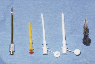
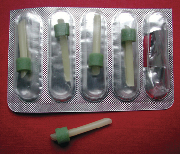
The best treatments for acute teat-end injury include symptomatic antiinflammatory therapy and reducing further trauma to the teat end. Each injury must be assessed individually. If milkout is simply reduced but not prevented, milkers sometimes use dilators of various types between milkings to stretch the sphincter muscles, thus allowing machine milkout. If milkout is difficult, it is best to avoid further machine milking and to utilize a teat cannula to effect milking twice daily when the other quarters are machine milked. If cannulas are used, the milker must exert extreme care to avoid exogenous inoculation of the teat cistern with microbes. Therefore the teat end must be cleaned gently, and alcohol must be applied before introducing the sterile cannula. Usually a disposable 1-in plastic cannula is used for this purpose. After complete milkout, the teat end is dipped as usual and a repeat dip performed in 10 minutes. Alternatively, some practitioners recommend indwelling plastic cannulas that may be capped between milkings. Several types are available commercially. In addition to facilitating milkout, these indwelling cannulas act as dilators that may reduce the possibility of streak canal adhesions or fibrosis. Teat dilators impregnated with dyes are not favored because they seem to induce chemical damage to the steak canal. However, many owners use such dilators anyway (Figure 8-17). Wax and silicone teat inserts that may retain patency with less iatrogenic mastitis are commercially available. The wax insert is recommended for initial use, but it disintegrates after several days. Insertion of silicone rubber inserts after the wax has disintegrated is recommended. The inserts have comparable efficacy and antibacterial properties. Both inserts are readily available in the United States. Alternatively, milk can be drained from the gland, intramammary antibiotics infused, a wax teat insert (Figure 8-18) placed in the teat, followed by icing and bandaging the teat with no further milkout for 2 to 3 days.
Nursing care is helpful but unfortunately is often not available on modern dairy farms. Soaking the injured teat with concentrated Epsom salts in a cup of warm water for 5 minutes twice daily helps reduce edema and inflammation. It is most important to avoid further trauma to the teat end and to minimize the risks for developing mastitis. Therefore avoidance of machine milking is indicated for at least several days whenever possible.
Problems that have been associated with teat-end lesions include crusts, necrosis, ulceration (Figure 8-19), and mastitis. All result in continued pain to the patient and mechanical interference with milkout as a result of scab or exudate buildup. Gentle soaking in warm dilute Betadine solution (Purdue Pharma, Cranbury, NJ) followed by removal of crusts or exudate aids complete milkout. Teat injury predisposes the cow to mastitis, particularly infections with gram-positive organisms.

Figure 8-19 Chronic teat-end injury with an ulcerative bed of granulation tissue ringed by crusted edges. This type of wound repeatedly produces a crusty scab that interferes with effective milkout and is an extremely common sequela to acute teat-end injuries.
Gradual return to normal milking is hoped for in 3 to 7 days following acute teat-end injury. Subacute or chronic injuries that continue to interfere with milkout may necessitate surgical intervention. Surgery should be avoided in acute teat-end injury because any sharp injury to an already damaged sphincter muscle and streak canal only serves to worsen the acute inflammation and hemorrhage, as well as the ensuing fibrosis. If milk flow is still obstructed after edema has resolved, examination should determine site of injury, fibrosis, or granulation tissue obstruction. Granulation tissue at the most dorsal aspect of the streak canal or most ventral part of the teat cistern is common. Fibrosis of the sphincter musculature also is very common. Instrument manipulation or sharp surgery on the teat end is then indicated. Wax inserts should be used to decrease stricture.
Before surgical intervention, the quarter should be full of milk. Experienced owners will not milk out the affected quarter before the veterinary visit, but if they have forgotten and done so, the cow should be given 20 units of oxytocin IV to fill the quarters. Without adequate milk in the quarter, it is impossible to assess how much the obstruction has been relieved.
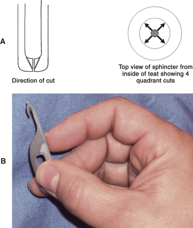
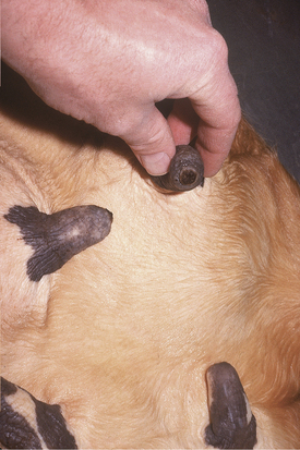
For surgical correction of obstructed teat ends, the teat should be washed, cleaned, and disinfected with alcohol (Box 8-1). The cow should be restrained and/or sedated before surgery. A teat bistoury or knife, preferably one with a small single cutting edge and blunt tip, should be used. The aim of this procedure is to relieve the stricture in the streak canal through two to four angled cuts made at 90-degree intervals (Figure 8-20, A and B). The cuts are made into the dorsal sphincter muscle but tapered so as not to cut the distal sphincter or teat end. We prefer the use of a Larsen teat blade because it allows a better control of the dept of the cut and facilitates the creation of a tapered incision. These radial incisions release the sphincter and frequently are the only treatments required. Some veterinarians use wax inserts to reduce hemorrhage following this procedure and to diminish subsequent inflammation and swelling that may impede milkout.
Box 8-1 Preparation for Teat Surgery or Treatment
Surgical thelotomy or repair of full-thickness lacerations and fistulae

Figure 8-20 A, Schematic illustration of teat knife incisions required to relieve sphincter muscle fibrosis. B, Preferred teat bistoury for radial cut for treatment of streak canal fibrosis. Note cutting edge is in the acute angle and thus allows control of the depth of the incision. In addition, if the operator flexes his/her wrist while pulling distally, only the proximal half of the streak canal will be incised.
A Moore’s teat dilator also has been used for sphincter muscle fibrosis. This instrument is inserted into the teat following routine preparation and advanced slowly to stretch the sphincter muscle without sharp surgery.
Masses of granulation tissue in the streak canal can act as an obstruction between the canal and teat cistern. They are generally a result of injury to the rosette of Furstenberg. These masses or growths are generally removed with the aid of a Hug’s teat tumor extractor. This instrument can be opened to allow excessive tissue to be grasped and cut off by the sharp edge of the extractor. It is a commonly used teat instrument, but care should be taken not to excise excessive surrounding healthy mucosa when removing granulation or fibrous tissue. The collateral mucosal damage associated with the blind use of the teat tumor extractor frequently results in recurrence of the stenosis. To precisely remove diseased tissue, and leave adjacent healthy tissue undisturbed, thelotomy with sharp incision is indicated (see teat-cistern obstructions). Minimally invasive fiberoptic theiloscopy in combination with electrosurgery is preferable, but the equipment is expensive, although it leads to improved long-term outcomes. The equipment is available from the Karl Storz Company (Charlton, MA).
Occasional instances of prolapsed streak canal mucosa are observed following crushing teat-end injuries. This tissue should be cut off flush with the teat end and then gently probed with a teat cannula to replace any everted tissue back into position in the streak canal.
Most veterinarians initially are too cautious and conservative when treating teat-end fibrosis. Experience is necessary to know “how much to cut” to allow not only short-term results but also to avoid subsequent reoperation because of recurrence of the problem. If in doubt, it is best to be conservative because the procedure always can be repeated. Most experienced veterinarians not only want to see a reduced resistance to hand milkout but also a slight dripping of milk immediately postoperatively. This dripping usually subsides as sphincter tone improves following resolution of dilatation associated with surgical instrumentation.
Repeated self-induced teat-end trauma to a specific teat dictates evaluation of the cause. Foot-induced trauma may be detected by smearing dye on the medial dewclaw and observing the teats for dye transmission onto the teat. In this case, removal of the medial dewclaw may help prevent injury in the future. Cows with pendulous udders that suffer repeated teat-end injuries usually have to be culled.
Teat-end necrosis or ulceration is difficult to manage because buildup of scab material in the crater-like ulcer recurrently interferes with milking. Gentle soaking and mechanical removal of the scabs are necessary for milkout. A mild teat dip with glycerin or lanolin for softening is indicated in these patients. Some require surgical manipulation if continued irritation or overmilking damages the sphincter muscle or dorsal streak canal. When teat-end necrosis is observed in more than one cow in a herd, the milking machinery and procedures should be examined carefully to rule out excessive vacuum pressure and physical or chemical irritants in teat dip or bedding (Figure 8-21).
Acquired Teat-Cistern Obstructions
Etiology
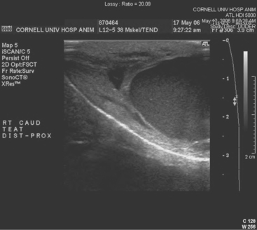
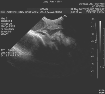
Teat-cistern lesions resulting in obstructed milk flow may be focal or diffuse. Most teat-cistern obstructions result from proliferative granulation tissue, mucosal injury, or fibrosis—all secondary to previous teat trauma. Occasional cases have no history of previous acute injury. Focal and diffuse lesions in the cistern cause an increasing degree of flow restriction that interferes with effective milk delivery to the streak canal during machine milking. With ultrasound examination, obstruction at the junction of the gland and teat cistern can be visualized (Figures 8-22 and 8-23). This type of obstruction leads to slow refill of the teat cistern such that they cannot be milked by machine. However, they can be milked by teat cannula or siphon. In addition to fixed lesions, floating objects known as “milk stones” or “floaters” may cause problems in milkout because they are pulled into the teat and mechanically interfere with milking. These floaters may be completely free or may be attached to the mucosa by a pedunculated stalk. Mucosal detachments also are encountered secondary to external teat trauma. The detached mucosa folds onto the opposite teat wall, causing a valve effect as milking progresses. Submucosal hemorrhage or edema from previous trauma is thought to cause detached mucosa; the problem may not be apparent until resolution of the submucosal fluid allows the detached mucosa to become mobile within the cistern.

Figure 8-22 Sonogram of the junction of the teat and gland cistern made with a convex 8.5-MHz probe (distal is to the left). At this location, the lumen abruptly narrows from 2 cm to 3 mm because the wall of the teat is thick and irregular. Also, a 3-mm thick band of tissue occludes the lumen of the teat cistern.
(Courtesy Dr. Amy Yeager, Cornell University.)

Stay updated, free articles. Join our Telegram channel

Full access? Get Clinical Tree

